The eLitMed.hu medical portal uses computer cookies for convenient operation. Detailed information can be found in the Cookie-policy.
Hungarian Radiology - 2007;81(05-06)
Content
Hungarian Radiology
OCTOBER 20, 2007
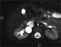
[Technical aspects of MR urography based on two cases]
[Intravenous urography has been the gold standard in diagnostic radiology studying the urogeintal system. However, ultrasound and other cross-sectional imaging methods have brought major change in this area. Beside modern (multislice) CT examinations the methods of MR urography are more frequently applied making possible both static and dynamic examinations beyond the wellknown advantages of MR imaging. The authors describe two complemetary methods of MR urography which provide a complete evaluation of the urinary system. The first method is aimed to image stationary fluid by using heavily T2 weighted turbo spin echo pulse sequences. This measurement well depicts the dilations of the excretory system even with little or no excretion of urine. The second method is analogous with the traditional IVU, as the excretion of a less nephrotoxic gadolinium based contrast medium followed by a T1 weighted gradient echo pulse sequence (possibly dynamic) helps to visualise the renal cavities and the urinary pathways. Normal renal function is a prerequisite when using this technique in order to visualise normal and obstructed urinary pathway disorders. The use of MR urography together with traditional MR methods may significantly reduce the number of invasive examinations and methods based on X-ray radiation exposure. MR urography may be exceptionally important in the uroradiological study of distensions of renal cavities in pediatric cases, pregnant women and renal transplant patients or in case of contrast material allergy.]


Hungarian Radiology
OCTOBER 20, 2007
[Pancreas transplantation: Indication, surgery, complications, diagnostic imaging and outcome]
[Diabetes mellitus is often leading to chronic microvascular and macrovascular complications. At present time, wholepancreas transplantation is the only option to achieve long-term insulin independence. Therefore, simultaneous kidney-pancreas transplantation has become the preferred treatment option for selected patients with type I diabetes mellitus and renal failure. In this review authors describe the indications and main types of pancreas transplantation: simultaneous pancreaskidney transplantation, renal transplantation first then pancreas transplantation, pancreas transplantation alone, islet-cell transplantation, and deal with surgical technical aspects. The immunological, metabolic, inflammatory, technical and late complications are also datailed as well as the outcome of the operations. The importance of the various imaging techniques is emphasized. The rate of mortality and complications are significantly higher after simultaneous kidney-pancreas transplantation than those after kidney transplantation alone. The role of the radiologist is to promote a successful outcome by early detection of the complications.]
Hungarian Radiology
OCTOBER 20, 2007
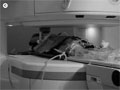
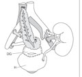
[MRI-guided prostate brachytherapy: First Hungarian experiences based on a canine study]
[INTRODUCTION - Modern radical radiotherapy can be an effective alternative of radical prostatectomy in low risk patients with prostate tumor. Our objective was to demonstrate the feasibility of transperineal MR-guided prostate interventions in an open MR unit and to present our early clinical experiences on canines. METHODS AND MATERIALS - The procedures were performed on 5 canines in an open-configuration 0.35T MR scanner. For interventions an MR compatible custom-made device was used. The canines were placed in the right lateral decubitus position. Template reconstruction, trajectory planning, target and OAR delineation were based on T2 FSE images. For image guidance and target confirmation, fast spoiled gradient-echo (FSPGR) sequence was used. MR compatible coaxial needles were inserted through the perineum to the base of the prostate. After satisfactory position was confirmed, brachytherapy catheters were placed through the coaxial needles, which were then removed. RESULTS - Mean and standard deviation of the needle displacements was 2.2 mm±1.2 mm, with a median of 2 mm. 96% of the errors were less than 4.0 mm. Implantation induced prostate motion was measured with a mean of 10.3 and 2.3 mm in cranio-caudal and transverse directions. Significant movement was only observed during the first 4 needle insertions. The average time needed for each step was: anesthesia 15 minutes, setup and positioning 15 minutes, initial imaging 15 minutes, template registration and projection 15 minutes, contouring, trajectory planning, insertion of 10 needles 60 minutes. CONCLUSION - Based on our canine model experiences our method seems to be a promising approach for performing feasible, accurate, reliable and high-quality prostate MR guidance within a reasonable time span. We plan to introduce MR-guided biopsy and brachytherapy in human patients in the near future.]
Hungarian Radiology
OCTOBER 20, 2007
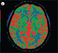
[Computed tomography brain perfusion in the management of acute stroke]
[INTRODUCTION - The multidetector CT-technology made the application of perfusion CT-examination in the diagnosis of vascular brain damages possible in recently. The purpose of this study was to introduce the method and to assess the importance of computed tomography brain perfusion in emergency patient care and early diagnosis of brain ischemia. PATIENTS AND METHODS - We perform brain perfusion examinations with a 2 slice multidetector computer tomography (General Electric Highspeed NX/i, 2004) in our hospital. We examined the results of native and perfusion CT of 27 patients who underwent CT brain perfusion examination during emergency patient care in our department between 2004 January and 2006 December. We also examined if the patients got systemic thrombolysation and the patients’ condition after therapy. RESULTS - The perfusion software can make quantitative colour maps of parameters (CBF, CBV, MTT) and can visualize mean value and percentil decrease of measuring parameters. There were 18 positive and 8 negative CTbrain perfusion examinations in the examined period. One examination was technically unvaluable. CONCLUSION - By measuring blood flow's decrease the CT-brain perfusion examination can separate the reversible and irreversible damage of brain parenchyma. The examination protocol of brain vascular damages are native CT-scan, postcontrast perfusion CT-examination and CTangiography by the recommendation of international literature. Despite the multidetector CT-s and CT-perfusion technic is available for years, the CT-brain perfusion examination is not a routine process in the emergency patient care in our country.]
Hungarian Radiology
OCTOBER 20, 2007

Hungarian Radiology
OCTOBER 20, 2007
[Radioguided localization of the non-palpable breast lesions and sentinel lymph nodes]
[INTRODUCTION - As the result of mammographic screening of breast cancer, the incidence of surgically removable, non-palpable breast lesions is increasing. The study is aimed to demonstrate the use of radioisotope and gamma-probe for identifying the sentinel lymph node and the intraoperative radioguided localization of occult breast lesions. PATIENTS AND METHODS - 26 patients with nonpalpable breast lesions underwent radioisotope guided sentinel lymh node localization for staging purposes. In eight patients non-palpable breast lesions were localized with use of radioisotope method intraoperatively. RESULTS - All breast lesions were successfully localized pre- and intraoperatively. The combined radioisotope and blue-dye staining method increased the sensitivity of the procedure for identifying sentinel lypmh node. In case of 14 patients with negative sentinel node, the surgical dissection of axillary lymph nodes was avoided. CONCLUSION - Radioguided localization is a simple, quick and accurate technique for localization of nonpalpable breast lesions and sentinel lymph node, which is utilized for the up-to-date and correct oncological management of the breast cancer patients.]
Hungarian Radiology
OCTOBER 20, 2007
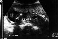
[Prenatal detection of campomelic dysplasia by sonography]
[INTRODUCTION - The campomelic dysplasia is a disorder characterized by short and bowed lower limbs resulting in dwarfism. CASE REPORT - In the case of a 21-year-old primipara woman the second screening ultrasonography raised the suspicion of short and bowed lower limbs of the fetus, at the 19th week of the pregnancy. Repeated examinations proved the presence of short and bowed femurs and tibias and abnormal echogenecity of the bones. The upper limbs were almost normal in length. During the 19th week of pregnancy, after a genetic analysis in agreement of the parents the pregnancy was interrupted without any complication. Photography and Xray of the fetus confirmed the diagnosis. CONCLUSION - Fetal ultrasonography should include exact size measurement and observation of the shape of the long bones, making possible the early detection of limb anomalies. The anomaly being proven by positive genetic analysis the pregnancy can be interrupted at the parents' request.]
Hungarian Radiology
OCTOBER 20, 2007
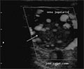
[Jugulotympanic glomus tumor - 7-year follow up of a rare disease]
[INTRODUCTION - The jugulotympanic glomus tumor is rare pathology with slow progression and its treatment is difficult. We present the imaging and clinical findings of a case obtained during 7 year follow up period. CASE REPORT - A 77-year-old female patient was admitted in 2000 with slowly progressing swelling of the neck. A palpable soft, non-painful resistance was noted at the main neck vessel region. Angiography revealed a jugulo-tympanic glomus tumor and embolization of the feeders from the external carotid artery was also performed. The size of the tumor is decreased but a clinically silent floating thrombus appeared in the internal jugular vein. It was treated with LMWH. Three years later external carotid artery transsection and denudation was performed. Slow progression of the tumor toward the subclavian vein was detected without intracranial invasion, in 2004. Repeated angiography was performed in 2005, but embolization was not possible due to technical reasons. Irradiation was considered to diminsh clinical symptoms, however due to the old age of the patients and the risk of bleeding, the treatment was not performed. Symptomatic therapy has been applied. CONCLUSION - Jugulo-tympanic glomus tumor is one of the non-chromaffin paragangliomas. The clinical symptomps are dominated by the paresis of IX.-X.-XI. nerves. Semimalignant disease and metastases are rare. The course is mostly influenced by compression syndromes and intracranial spread. The therapy is mostly microsurgical but preoperative embolization is frequently done to decrease blood loss. Surgical therapy is recommended with prior embolization of the feeders in order to decrease the blood supply of the mass. If surgery cannot be carried out irradiation therapy is needed. Unfortunately, all of these procedures are seldom curatives because of natural course of the disorder and the high frequency of recurrencies.]
Hungarian Radiology
OCTOBER 20, 2007
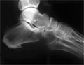
[Tarsal coalitions in our adult patients]
[INTRODUCTION - Tarsal coalition is an abnormal bony, cartilaginous or fibrous union between two or three bones of the mid and hindfoot. It occurs mostly (90%) in the calcaneonavicular and talocalcaneal joints. Coalitions are usually detected in the second decade of life, but several cases are noted in children and in later adulthood. Estimated incidence is 1-2% of the total population. CASE REPORTS - Cases of unilateral tarsal coalitions (five calcaneonavicular, three talocalcaneal) in four male and three female patients are presented. The average age of the patients is 30 years. The radiologic diagnosis was based on the secondary signs seen on the lateral plain films of the feet. These signs are parallel with clinical symptoms, in contrary to the primary radiologic signs. The two types of the anomaly coexisted in a female patient. CONCLUSION - Routine lateral not-magnified pedal radiographs are valuable screening tool for tarsal coalitions.]
Hungarian Radiology
OCTOBER 20, 2007

Hungarian Radiology
OCTOBER 20, 2007

Hungarian Radiology
OCTOBER 20, 2007

Hungarian Radiology
OCTOBER 20, 2007

Hungarian Radiology
OCTOBER 20, 2007

Hungarian Radiology
OCTOBER 20, 2007

Hungarian Radiology
OCTOBER 20, 2007

1.
Clinical Neuroscience
[Headache registry in Szeged: Experiences regarding to migraine patients]
21. MAY
2.
Clinical Neuroscience
[The new target population of stroke awareness campaign: Kindergarten students ]
21. MAY
3.
Clinical Neuroscience
Is there any difference in mortality rates of atrial fibrillation detected before or after ischemic stroke?
27. NOV
4.
Clinical Neuroscience
Factors influencing the level of stigma in Parkinson’s disease in western Turkey
27. SEP
5.
Clinical Neuroscience
[The effects of demographic and clinical factors on the severity of poststroke aphasia]
18. JUL
1.
2.
3.
4.
5.