The eLitMed.hu medical portal uses computer cookies for convenient operation. Detailed information can be found in the Cookie-policy.
Hungarian Radiology - 2007;81(01-02)
Content
Hungarian Radiology
MARCH 20, 2007
[Diagnostic and therapeutical possibilities in constipation]
[Constipation is a common gastrointestinal problem. The prevalence of symptoms related with constipation fluctuates from 3 to 20 per cent. Constipation occurs more frequently in the elderly people and in females and more frequent in case of inactivity and less fiber intake. Assesment of patients with severe constipation includes specialized investigations. Exclusion of primary organic causes has to be the first step, then metabolic, neurological and iatrogenic causes (such as medicament side effects, etc) have to be excluded. After these considerations special functional gastroenterological investigations are needed which contribute to the diagnosis and differential diagnosis of the cause of the constipation. Anorectal manometry, ballon expulsion test, defecography and colon transit studies allow us to distinguish between slow colon transit, colon inertia, different subtypes of outlet obstruction, and the constipation predominant irritable bowel syndrome. The evaluation of these specific studies leads to the exact diagnosis and appropriate treatement for their problem can be given to the patients, which always has to be individually planed in all cases.]


Hungarian Radiology
MARCH 20, 2007

[The radiohygienic aspects of the interventional radiology]
[Interventional radiology is a relatively new and very rapidly developing cost-effective branch of radiology. Its aim to help or to replace surgical procedures and interventions in many cases are life saving, which are performed by imaging modality control (most commonly angiography or fluoroscopy). During interventional radiological procedures the exposure of staff and patients is usually higher, than in conventional radiography or fluoroscopy. Deterministic effects may also occur. The dosimetry can be carried out by film dosimetry, thermoluminescent dosimetry, DAP meters, semiconductor detectors and personal electronic dosimeter. The basis of reduction of radiation exposure is the radiation protection training. An important rule is that reduction of patient exposure is connected with reduction of staff exposure. With the use of appropriate tools and training the most injuries are avoidable.]
Hungarian Radiology
MARCH 20, 2007

Hungarian Radiology
MARCH 20, 2007

[Teleradiology - opportunity or threat?]
[Teleradiology - as a result of recent developments in digital imaging and informatics - appears to be a technology potentially responding to many challenges in the field of diagnostic radiology. It may help in the centralization of service, in the support of emergency care, and in the more accurate diagnosis of cases requiring special skill. Outsourcing of imaging diagnostic reporting activities may solve human resource problems and may decrease wage expenditures. Nevertheless teleradiology exposes also some difficulties (human and technical aspects) which we should recognize in time for being able to protect ourselves.]
Hungarian Radiology
MARCH 20, 2007
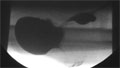
[Transcranial Doppler monitoring of distal embolism during of carotid stenting]
[INTRODUCTION - Reducing the risk of embolisation during endovascular treatment of internal carotid artery stenosis is very important. The rate of embolisation is affected by the different steps of stenting manipulation. Using transcranial Doppler equipment we studied the embolic signals during the different phases of carotid dilatation and stenting. MATERIAL AND METHOD - 50 patients (33 male, 17 female; mean age 64 years) were intraproceduraly monitorized with transcranial Doppler. Predilatation was necessary in nine cases, postdilatation was performed in 39 cases. The number of emboli were measured in seven different steps of endovascular treatment of carotid stenosis. Different type of commercial available endovascular devices were used. RESULTS - Intraprocedural embolisation was observed in every case. In different phases of carotid stenting the rate of embolisation showed marked differences in each phase of carotid stenting. Crossing the stenosis with stent delivery system were accompanied by a low rate of embolism (5.3) compared to the level during stent opening (9.16) and balloon dilatation (9.96). The highest level of embolisation was observed during predilatation (15.9) without the protection of the stent. CONCLUSIONS - We detected embolisation in all of the cases, however the number of embolic signals varied in different phases of carotid artery stenting. Embolisation can be reduced if the most dangerous steps (i.e. pre- and postdilatation) are avoided. Using TCD monitorisation the physician can be informed by the degree of embolisation that may alarm the interventionalist to perform the procedure more carefully, furthermore it can be employed during the training of carotid stenting.]
Hungarian Radiology
MARCH 20, 2007

Hungarian Radiology
MARCH 20, 2007
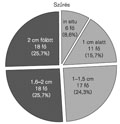
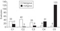
[Results of breast cancer screening and clinical mammography at the Kenezy Breast Center, Debrecen between 2002-2003]
[INTRODUCTION - Breast cancer screening has been started in January 1. 2002. in Hungary in the course of the National Health Program. Breast cancer is the main cause of death among women’s malignant tomors, and the aim of the project is to reduce this mortality. The chance of survival is highly increased by the early detection of the disease. Kenezy Breast Center was connected to this project. PATIENTS AND METHODES - Females between 45-65 years without symptoms participated in the project. Paralel to this women with symptoms, sometimes with palplable masses were clinically examined. Screening mammography films were read by two radiologists and the complementary examinations of the breast and the axillary lymph nodes - ultrasonography, guided biopsy (FNAB, core biopsy) - were performed always by the same doctor. Results of the two projects were compared. RESULTS - The incidence of malignant breast cancer was 4‰ in the screening and 1,5% in the clinical group. 46.5% of the malignant breast cancers revealed by the clinical examinations was diagnosed in the group of women between the age of 45 to 65 years. This is the age when most women are involved in the screening program. 7.3% of the tumors was diagnosed in the 40- 44 year age-group and 11.3% among women aging 66-77 years. The rate of malignant tumors smaller than 1.5 cm was 49.1% according to screening records and 36% in the clinical trial. In both groups, tumor size of 1.5 cm proved to be a critical limit regarding to the development of metastases, mainly in the axillary region. Above this size, metastases were more frequent. CONCLUSIONS - Both breast screening program and clinical exams are of great significance. Based on the data obtained during two years, authors found that women below the age of 40 and above the age of 65 should also be involved in the screening program. Detection of breast tumor is possible at an early stage by screening. In the case of small tumors (smaller than 1.5 cm) the development of axillary metastases is less likely than in the case of larger ones. The lack of metastases in the axillary lymph nodes offers better prognosis according to the published scientific data, which reinforces the importance and necessity of the screening programs.]
Hungarian Radiology
MARCH 20, 2007
[Az emlődaganatok radiológiai vizsgálatának újdonságai Onco Update, 2007]
[Experiences about the breast diagnostic methods are accumulating year-to-year, rapidly. Therefore the current examination algorithm is changing continuously. New diagnostic and therapeutic modalities are entering into the daily practice. Some of them became obsolete, so far their application is becoming a faulty decision. Some other methods become obligatory steps in the diagnostics. These are the reasons why the up-to-date knowledge of the literature is mandatory. Systematic review of the most recent articles of the last two years (January 2005-December 2006) of breast radiological diagnostics and the actual place of the imaging and interventional methods are presented. The following topics are summarized: breast cancer screening with conventional and digital mammography, computer assisted diagnostics (CAD), high risk patients' screening, US, MRI, MSCT, PET/CT, diagnostic interventions, differential diagnostics, percutaneous tumour ablation, therapy-related questions in the diagnostic work up.]
Hungarian Radiology
MARCH 20, 2007
Hungarian Radiology
MARCH 20, 2007

Hungarian Radiology
MARCH 20, 2007

Hungarian Radiology
MARCH 20, 2007

Hungarian Radiology
MARCH 20, 2007

Hungarian Radiology
MARCH 20, 2007
Hungarian Radiology
MARCH 20, 2007

Hungarian Radiology
MARCH 20, 2007

Hungarian Radiology
MARCH 20, 2007

Hungarian Radiology
MARCH 20, 2007

Hungarian Radiology
MARCH 20, 2007

1.
Clinical Neuroscience
[Headache registry in Szeged: Experiences regarding to migraine patients]
21. MAY
2.
Clinical Neuroscience
[The new target population of stroke awareness campaign: Kindergarten students ]
21. MAY
3.
Clinical Neuroscience
Is there any difference in mortality rates of atrial fibrillation detected before or after ischemic stroke?
27. NOV
4.
Clinical Neuroscience
Factors influencing the level of stigma in Parkinson’s disease in western Turkey
27. SEP
5.
Clinical Neuroscience
[The effects of demographic and clinical factors on the severity of poststroke aphasia]
18. JUL
1.
2.
Clinical Oncology
[Pancreatic cancer: ESMO Clinical Practice Guideline for diagnosis, treatment and follow-up]
29. AUG
3.
Clinical Oncology
[Pharmacovigilance landscape – Lessons from the past and opportunities for future]
29. AUG
4.
5.