REM sleep, REM parasomnias, REM sleep behaviour disorder
SZÛCS Anna 1, MUTTI Carlotta2, PAPP Anikó3, HALÁSZ Péter3, PARRINO Liborio 2
MAY 30, 2022
Clinical Neuroscience - 2022;75(05-06)
DOI: https://doi.org/10.18071/isz.75.0171
Review
SZÛCS Anna 1, MUTTI Carlotta2, PAPP Anikó3, HALÁSZ Péter3, PARRINO Liborio 2
MAY 30, 2022
Clinical Neuroscience - 2022;75(05-06)
DOI: https://doi.org/10.18071/isz.75.0171
Review
Szöveg nagyítása:
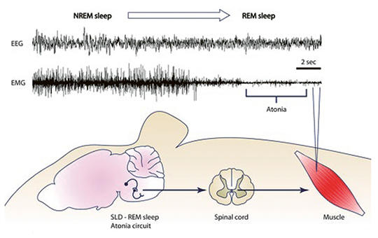
We review the literature on REM parasomnias, and their the underlying mechanisms. Several REM parasomnias are consistent with sleep dissociations, where certain elements of the REM sleep pattern emerge in an inadequate time (sleep paralysis, hypnagogic hallucinations and cataplexy) or are absent/partial in their normal REM sleep time (REM sleep without atonia, underlying REM sleep behavior disorder). The rest of REM parasomnias (sleep related painful erection, catathrenia) may have other still unclear mechanisms. REM parasomnias deserve attention, because in addition to disturbing sleep and causing injuries, they may shed light on REM sleep functions as well as the heterogeneous etiologies of parasomnias. One of them, REM sleep behavior disorder has special importance as a warning sign of evolving neurodegenerative conditions mainly synucleinopathies (some cases synucleinopathies themselves) and it is a model parasomnia revealing that parasomnias may have by autoimmune, iatrogenic and even psychosomatic etiologies.
Clinical Neuroscience
Over the past year, many cases with newly onset or significantly exacerbated tic disorders were observed worldwide, where some aspects of the clinical presentation or the symptomatology were atypical for established tic diagnoses. Our purpose was to describe the atypical cases and raise relevant diagnostic issues. Consecutive cases with atypical tic presentations were documented. Five atypical tic cases are described. These cases shared some common characteristics, most notably the fact that all of them had been exposed to online presentation of ticking behaviour on social media platforms prior to the de novo development or exacerbation of their tics. Even though the order of events suggests causality and therefore the diagnosis of a functional tic disorder, unambiguous criteria for classifying atypical tics as functional symptoms are lacking. Differentiating neurodevelopmental and functional tics in childhood is currently problematic. Based on the currently unresolved issues in differential diagnosis, the importance of watchful waiting and behavioural interventions is highlighted to avoid unwarranted pharmacotherapy.
Clinical Neuroscience
Prevalence of acute ischemic stroke (AIS) is increased in patients with coronavirus disease 2019 (COVID-19). A proposed hypothesis is increased virus-induced propensity to hypercoagulation resulting in arterial thrombosis. Our aim was to provide evidence regarding the involvement of neutrophil extracellular trap (NET) formation (NETosis) in COVID-19 related AIS. Twenty-six consecutively enrolled COVID-19+ pneumonia patients with AIS, 32 COVID-19+ pneumonia patients without AIS and 24 AIS patients without COVID-19 infection were included to the study. Clinical characteristics of recruited patients were collected. Serum levels of citrullinated histone H3 (H3Cit; a factor of NETosis), IL-8 and C5a (mediators associated with NETosis) were measured by ELISA (enzyme-linked immunosorbent assay). H3Cit levels were significantly higher in COVID-19+ AIS patients, whereas all study groups showed comparable IL-8 and C5a levels. There were no significant differences among etiological subgroups of AIS patients with or without COVID-19. AIS patients with COVID-19 showed relatively increased white blood cell, lymphocyte, neutrophil, D-dimer, C-reactive protein and procalcitonin levels than control groups. H3Cit levels did not correlate with clinical/prognostic features and inflammation parameters. H3Cit and IL-8 levels were correlated in COVID-19 patients without stroke but not in COVID-19 positive or negative AIS patients. Increased levels of inflammation parameters and H3Cit in COVID-19 related AIS suggest that NETosis may cause susceptibility to arterial thrombosis. However, H3Cit levels do not correlate with clinical severity measures and inflammation parameters diminishing the prognostic biomarker value of NETosis factors. Moreover, the link between IL-8 and NETosis appears to be abolished in AIS.
Clinical Neuroscience
Neurological symptoms and complications associated with coronavirus 2019 (COVID-19) are well known. It was aimed to evaluate the brainstem and trigeminal/facial nerves and the pathways between these structures in COVID-19 using the blink reflex test. Thirty patients with post COVID-19 (16 males, 14 females) and 30 healthy individuals (17 males, 13 females) were included in this prospective study. Individuals who previously had a positive nose swap polymerase chain reaction test for severe acute respiratory syndrome coronavirus 2 and whose previously clinical features were compatible with COVID-19 were included in the post COVID-19 patient group. Neurological examination of the participants should be normal. Blink reflex test was performed on all participants. R1, ipsilateral R2 (IR2), and contralateral R2 (CR2) waves obtained from the test were analyzed. The mean ages of healthy individuals and post COVID-19 patients were 34.0±6.4 and 38.4±10.6 years, respectively. Both age and gender were matched between the groups. R1, IR2, and CR2 latencies/amplitudes were not different between the two groups. The side-to-side R1 latency difference was 0.5±0.3 and 1.0±0.8 ms in healthy individuals and post COVID-19 patients, respectively (p=0.011). One healthy individual and 12 patients with post COVID-19 had at least one abnormal blink reflex parameter (p=0.001). This study showed that COVID-19 may cause subclinical abnormalities in the blink reflex, which includes the trigeminal nerve, the seventh nerve, the brainstem, and pathways between these structures.
Clinical Neuroscience
Although severe acute respiratory syndrome coronavirus-2 (SARS-CoV-2) is a novel virus, many central and peripheral nervous system manifestations associated with coronavirus disease-19 (COVID-19) infection have been reported. Beyond the neurologic manifestations, we may still have much to learn about the neuropathologic mechanism of SARS-CoV-2 infection. Here we report a case of post-poliomyelitis syndrome (PPS) related to COVID-19 and attempt to predict the possible pathophysiologic mechanism behind this association.
Clinical Neuroscience
[Research results in recent years have demonstrated that B-lymphocytes play a crucial role in the pathogenesis of multiple sclerosis (MS). The increased understanding of the disease process has resulted in the development of B cell-targeting antibodies as potential drugs for both relapsing and progressive forms of MS. Therefore, B-cell depletion therapies are becoming more prominent and determining in reducing disease progression. The first B-cell depleting anti-CD20 monoclonal antibody was rituximab, which has also been studied in MS and, following favourable results, new drugs have been developed with a similar point of attack. In 2017, the FDA and in 2018, the EMA approved ocrelizumab, another anti-CD20 monoclonal antibody, for the treatment of relapsing-remitting (RRMS) and primary progressive multiple sclerosis (PPMS). This was a particularly significant advance in the treatment of PPMS, as it was the first medication with a proven effect of reducing progression in PPMS. Ofatumumab, a fully human anti-CD20 monoclonal antibody, has emerged recently as a new player in B-cell depletion therapy. The drug has also recently been approved by the EMA in March 2021 for use in relapsing forms of MS. In this review, we detail the mechanism of action and efficacy of anti-CD20 therapies currently used in MS. ]
Clinical Neuroscience
Natural disasters, such as earthquakes, frequently result in mood disorders among affected individuals. It is established that neuropathic pain arising from traumatic neuropathies is also linked to mood disorders. This study investigates the influence of neuropathic pain on the development of mood disorders in earthquake survivors with peripheral nerve injuries, following the earthquake centered in Kahramanmaraş on February 6, 2023.
Clinical Neuroscience
The aim of this study is to comprehensively determine the types of affected fibers in Parkinson’s disease (PD) patients by employing nerve conduction studies (NCS), sympathetic skin response (SSR) examinations, and current perception threshold (CPT) testing and to analyze the correlation between levodopa use and nerve involvement. This retrospective study included 36 clinically diagnosed PD patients who were recruited between January 2018 and April 2019.
Clinical Neuroscience
Parkinson’s disease (PD) is a heterogeneous neurodegenerative disorder characterized by contradictory clinical outcomes among its several subtypes. The disease can manifest with a tremor-dominant (TD) or a non-tremor-dominant (NTD) phenotype. Although the TD subtype may show a better prognosis, there is limited information on the phenotypic differences regarding the level of axial symptoms. For this reason, in this study it was aimed to make a quantitative comparison of axial posture and spinal mobility between PD with TD and NTD.
Journal of Nursing Theory and Practice
[The high blood pressure disease is one of the primary risk factors for the development of other cardiovascular diseases. The aim of the present study was to assess the medication habits, sleep quality, and quality of life of patients aged 45 and older living with hypertension.
Our study was a quantitative descriptive cross-sectional survey conducted in 2022 in the form of an online questionnaire among patients aged 45 and older suffering from hypertension (n=143). The data collection tools included the Morisky Medication Adherence Scale (8 items), the Pittsburgh Sleep Quality Index, EQ-5D-5L, and a self-designed questionnaire.
Lower age was associated with higher levels of medication adherence (R=-0.36; p=0.024). Patients who regularly visited their general practitioner showed more consistent medication intake (p=0.048). Adequate sleep quality positively influenced the quality of life (p<0.001).
Regular visits to the general practitioner not only facilitate medication adherence but also provide an excellent opportunity for patient education and health promotion. Therefore, it has paramount importance to draw the attention of hypertensive patients to the role of regular check-ups with their general practitioner, which can contribute to the prevention of complications arising from hypertension.]
Clinical Neuroscience
Ciprofloxacin (CIP) is a broad-spectrum antibiotic widely used in clinical practice to treat musculoskeletal infections. Fluoroquinolone-induced neurotoxic adverse events have been reported in a few case reports, all the preclinical studies on its neuropsychiatric side effects involved only healthy animals. This study firstly investigated the behavioral effects of CIP in an osteoarthritis rat model with joint destruction and pain.
Effect of COVID-19 on seizures and patient behavior in people with epilepsy
Statistical evaluation of measured biomechanical properties of human brain aneurysm samples
1.
Clinical Neuroscience
Is there any difference in mortality rates of atrial fibrillation detected before or after ischemic stroke?2.
Clinical Neuroscience
Factors influencing the level of stigma in Parkinson’s disease in western Turkey3.
Clinical Neuroscience
Neuropathic pain and mood disorders in earthquake survivors with peripheral nerve injuries4.
Journal of Nursing Theory and Practice
[Correlations of Sarcopenia, Frailty, Falls and Social Isolation – A Literature Review in the Light of Swedish Statistics]5.
Clinical Neuroscience
[Comparison of pain intensity measurements among patients with low-back pain]1.
2.
Clinical Neuroscience Proceedings
[A Magyar Stroke Társaság XVIII. Kongresszusa és a Magyar Neuroszonológiai Társaság XV. Konferenciája. Absztraktfüzet]3.
4.
Journal of Nursing Theory and Practice
[A selection of the entries submitted to the literary contest "Honorable mission: the joys and challenges of our profession" ]5.
Journal of Nursing Theory and Practice
[End of Life and Palliative Care of Newborns in the Nursing Context]


COMMENTS
0 comments