The eLitMed.hu medical portal uses computer cookies for convenient operation. Detailed information can be found in the Cookie-policy.
Clinical Neuroscience - 2009;62(03-04)
Content
Clinical Neuroscience
MARCH 25, 2009

[Account on the 17. Congress of the Hungarian Society of Neuroradiology and on the position of neuroradiology in Hungary today]
[Account on the 17. Congress of the Hungarian Society of Neuroradiology and on the position of neuroradiology in Hungary today 2009;62(03-04)]


Clinical Neuroscience
MARCH 25, 2009
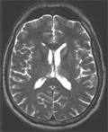
[MR imaging of acute disseminated encephalomyelitis and multiple sclerosis in children. A review (in English language)]
[Inflammatory diseases of the central nervous system (CNS) are relatively rare in children, but their relevance to public health is considerable due to frequent and significant long term morbidity and even mortality. As in adults, acute disseminated encephalomyelitis (ADEM) and multiple sclerosis (MS) and their variants are the most common entities in this group of pathologies in the pediatric patient population. Recent efforts have focused on establishing standardized diagnostic criteria schemes to facilitate the diagnosis and differential diagnosis of these diseases, however especially with multiple sclerosis those have not been fully validated yet for disease occurring in children. In recent decades the role of MRI has been constantly increasing in the diagnostic work-up of suspected inflammatory diseases of the CNS as well as in the follow-up of patients with confirmed disease. Currently, MRI is the first-line diagnostic imaging modality in ADEM and MS and is fully integrated in the most widely used diagnostic criteria schemes, but it has a key role in clinical therapeutic research trials as well. This paper provides an update on the current concepts and strategies of MRI in inflammatory diseases of the CNS, as well as a review of the imaging semiology of the various disease entities and variants with emphasis on clinical and imaging particularities relevant to the pediatric patient population.]
Clinical Neuroscience
MARCH 25, 2009
[Current issues of neuroimaging diagnostics of multiple sclerosis]
[Magnetic resonance imaging techniques have been routinely used in diagnostics and follow-up of multiple sclerosis and magnetic resonance imaging findings are incorporated into the current diagnostic criteria of the disease. International guidelines were created aiming to define the indication, timing, minimum requirements and interpretation of magnetic resonance examination in multiple sclerosis. In Hungary, there is a lack of widely-accepted standardized protocol, therefore presenting the magnetic resonance diagnostic criteria and the international guidelines may prove useful. It may also point towards consideration of a national guideline.]
Clinical Neuroscience
MARCH 25, 2009
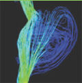
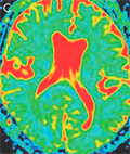
[Application of diffusion weighted imaging in neuroradiology]
[Diffusion weighted magnetic resonance (MR) imaging is available on all modern MR scanners. It depicts the motion of water molecules in the brain tissue and intracranial foreign tissues and provides information on changes in the proportion of intra- and extracellular space and the characteristics of foreign intracranial tissues and fluids. It is of utmost importance eg. in the diagnostics and differential diagnostics of acute ischaemic brain lesions, the diagnostics of inflammatory brain processes and in the differential diagnostics of intracranial space-occupying lesions. The examination method of short scanning and post-processing time must be completed with the apparent diffusion coefficient (ADC) maps and it is indispensable in the everyday neuroradiological diagnostics. Diffusion tensor imaging and tractography are able to depict the white matter tracts. They require a longer scanning and post-processing time and have several technical problems yet to be solved, but they provide help in their current state e.g. in the surgical planning of intracranial space-occupying lesions.]
Clinical Neuroscience
MARCH 25, 2009
[State of the art neurointerventional treatment of intracranial vascular anomalies]
[Haemorrhagic stroke is frequently caused by rupture of intracranial vascular anomalies. The role of minimally invasive therapy in the treatment of such lesions has increased dramatically within the past two decades. The purpose of this study is to summarize the pathology and clinical features of these anomalies and to overview the potential applications of neurointerventional techniques in their treatment. Endovascular therapy is the first choice of treatment for most intracranial aneurysms. Both pial and dural arteriovenous malformations are being treated by endovascular techniques, but the combination of different modalities (such as endovascular, direct surgery and radiosurgery) is frequently applied. Capillary malformations require surgical removal and venous anomalies do not allow for any type of invasive treatment. State of the art therapy of intracranial vascular anomalies require institutions equipped with appropriate imaging facilities and having equal access to both conventional neurosurgical and neurointerventional techniques with ample experience and case load.]
Clinical Neuroscience
MARCH 25, 2009
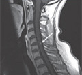
[Noninvasive imaging in the diagnosis of cerebral sinus thrombosis]
[Cerebral venous thrombosis is a severe, but potentially reversible disease, when it is promptly recognised and treated. Due to its varied and many aetiological factors and clinical manifestations, non-invasive radiological imaging plays a key role in the diagnostic procedure. Unenhanced and contrast-enhanced computer tomography (CT) and magnetic resonance (MR) imaging, CT angiography (CTA), MR angiography - such as time-of-flight (TOF) and phase-contrast (PC) angiography - are appropriate techniques for detecting cerebral venous flow and brain parenchymal changes. To achive adequate diagnosis timely it is necessary to have a correct knowledge of the venographic techniques, the temporally altering appearance of the thrombus, and the differential diagnostic problems that may occur. In our article, we summarized these characteristics by recent international publications and our own clinical observations and propose recommendations regarding the examination protocol of the dural sinus thrombosis.]
Clinical Neuroscience
MARCH 25, 2009
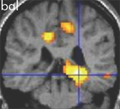
[Plasticity of brain lateralization in epilepsy: fMRI studies]
[Aims - This review is a summary of four studies investigating the effect of epileptic disturbance and other functional factors on the brain lateralization. Methods - Patients were investigated by functional MRI by using visual, sensorimotor, memory, and speech paradigms. Results - 1. Malformations of cortical development localized within speech centers induce contralateral speech reorganization in only 50% of cases. 2. Epileptic disturbance can induce a reorganization in language lateralization independent of the effect of lesion. 3. Bilateral epileptic activity can prevent a reorganization of memory functions contralateral to the seizure onset region. 4. Injury of the right upper limb can induce a left-to-right shift of language lateralization. Conclusions - Our results may have implication during neurorehabilitation process and neurosurgical interventions. Participation of malformations of cortical development in cognitive functions should keep in mind during surgery. Reorganization of speech- and memory functions should also be considered during operations of central nervous system.]
Clinical Neuroscience
MARCH 25, 2009
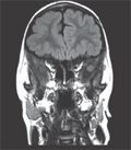
[Clonazepam can facilitate sensorimotor functional MRI examinations in status epilepticus during sleep]
[Functional magnetic resonance imaging examinations became an integral part of epilepsy surgery workup. In the pediatric population these examinations are usually carried out in full anesthesia, however in some forms of epilepsy, e.g. electrical status epilepticus in sleep, anesthesia could jeopardize the success of the examination. Here, we show on the example of an eight-year-old polymicrogyric epileptic child, that clonazepam can help suppressing the epileptic activity during propofol anesthesia, so it could facilitate performing passive sensorimotor functional MRI in such a case. Furthermore, among the methodological issues addressed, this case provides evidence for a post-hemispherotomy like functional redistribution of sensorimotor activations to the unaffected hemisphere in a case of childhood epilepsy.]
Clinical Neuroscience
MARCH 25, 2009

[Interhemispheric asymmetry of diffusion parameters in the human brain]
[Our aim was to study the hemispheric asymmetry of the human brain through the simultaneous measurements of the apparent diffusion coefficient (ADC) and fractional anisotropy (FA) values within a homogeneous sample in regards to the gender, age and handedness. Method - Eleven young women were included in our diffusion study performed on a 1.5 T MR scanner. We calculated the ADC and FA values in nine-nine region of interest points of the brain. The diffusion values were measured symmetrically in the right and left hemisphere of the brain and were then statistically compared to detect interhemispheric asymmetry. Results - We did not find any significant differences between the data of the two hemispheres in regards to the ADC values. However, we detected a significant right sided asymmetry in case of the globus pallidus in FA values (p=0.04). Conclusion - Our results of our study of young women suggest that diffusion asymmetry does not appear in case of ADC values and only slightly (in one ROI in our study) in FA values.]
Clinical Neuroscience
MARCH 25, 2009
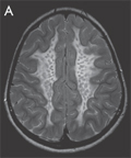
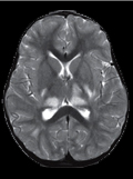
[Diffusion weighted magnetic resonance imaging of metachromatic leukodystrophy - a case report]
[We present the case of a 7 years old boy with the juvenile form of metachromatic leukodystrophy. Beside conventional sequences, a diffusion-weighted magnetic resonance study was performed. On T2-weighted images a diffuse hyperintensity of the cerebral white matter with a typical tigroid pattern was seen as a well-known characteristics of the disease. No difference was detectable on T2-weighted images between the different cerebral white matter regions. Diffusion study showed low apparent diffusion coefficient values in the temporal and orbitofrontal regions and normal values in other cerebral regions compared to the normal appearing cerebellar white matter, wich is partly consistent with the previously reported data. The results of early diffusion studies of the disease are not straightforward and the physical and biochemical backgrounds of the diffusion properties still remain unknown. The more data we collect, the closer we get to the understaning of the diffusion changes in metachromatic leukodystrophy.]
Clinical Neuroscience
MARCH 25, 2009

1.
Clinical Neuroscience
[Headache registry in Szeged: Experiences regarding to migraine patients]
21. MAY
2.
Clinical Neuroscience
[The new target population of stroke awareness campaign: Kindergarten students ]
21. MAY
3.
Clinical Neuroscience
Is there any difference in mortality rates of atrial fibrillation detected before or after ischemic stroke?
27. NOV
4.
Clinical Neuroscience
Factors influencing the level of stigma in Parkinson’s disease in western Turkey
27. SEP
5.
Clinical Neuroscience
[The effects of demographic and clinical factors on the severity of poststroke aphasia]
18. JUL
1.
2.
Clinical Oncology
[Pancreatic cancer: ESMO Clinical Practice Guideline for diagnosis, treatment and follow-up]
29. AUG
3.
Clinical Oncology
[Pharmacovigilance landscape – Lessons from the past and opportunities for future]
29. AUG
4.
5.