The eLitMed.hu medical portal uses computer cookies for convenient operation. Detailed information can be found in the Cookie-policy.
Clinical Neuroscience - 2013;66(09-10)
Content
Clinical Neuroscience
OCTOBER 05, 2013


Clinical Neuroscience
OCTOBER 05, 2013

[The impact of the vitamin D in neurological diseases and neurorehabilitation: from demencia to multiple sclerosis. Part I: The role of the vitamin D in the prevetion and treatment of multiple sclerosis]
[The world-wide incidence of vitamin D deficiency is high, independently of age. Multiple sclerosis is a chronic disorder, occuring in those who possess or are exposed to a combination of genetic and environmental risk factors. One of the environmental factors associated with the development is vitamin D. Vitamin D is an immunomodulatory agent, its role is verified in many of autoimmune diseases. Vitamin D inhibits IL-6, IL-17 and IL-23 secretions which are crucial in Th1 and Th17 differentiation and also decreases proinflammatorical cytokine production. Moreover it enhances the immunosuppressive IL-10 cytokine secretion and inhibits the T-reg cell development. These cytokines and cells are essential for the pathomechanism of multiple sclerosis. Data have shown, that the vitamin D levels above 100 nmol/l (40 ng/ml) is essential for the prevention of multiple sclerosis. Below this level the vitamin D supplementation is reasonable. In pregnancy, the vitamin D deficiency at the last two semester increases the risk for the multiple sclerosis of the infant. The optimal vitamin D level for multiple sclerosis patients is 100-150 nmol/l (40-60 ng/ml). There is no consensus for the role of vitamin D in multiple sclerosis yet, but until the achieving this, the diagnosis and the treatment of the vitamin D deficiency is crucial for scelrosis multiplex patients and in cases of elevated risk. Data shows, that in patient with multiple sclerosis the normal vitamin D level is suboptimal, however the exact role of vitamin D and doses must be clarified by interventional studies.]
Clinical Neuroscience
OCTOBER 05, 2013
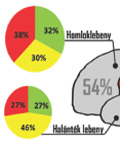
[Occurence and molecular pathology of low grade gliomas]
[Background - The WHO grade I. and II. low-grade gliomas represent nearly the 15% of all primary brain tumors. These tumours contain clinically, hisologically and molecularly distinct tumor types. According to their histologic characteristic, grade II glial tumours are the diffuse astrocytoma, oligodendroglioma and oligoastrocytoma subgroups; the ependymal tumors are not included in this study. Methods - In our publication, we analysed the histological diagnosed glioma cases between 2007 and 2011 at our institution. Results - Low-grade gliomas were diagnosed in 127 cases (62 male / 65 female), and the mean ages were 39 years (±20.3). More than half of the cancers were localizated in the frontal lobe, and the second most frequent area was the temporal lobe. Finally, we comlete our report with an overview of major molecular pathways in low-grade gliomas.]
Clinical Neuroscience
OCTOBER 05, 2013
[Occurence and molecular pathology of high grade gliomas]
[Background - Glial tumours represent the most frequent type of primary brain cancers. Gliomas are characterized by heterogeneity that makes the diagnosis, histological classification and the choosing of correct therapy more difficult. Despite the advances in developing therapeutic strategies patients with malignant gliomas have a poor prognosis; therefore glial tumours represent one of the most important areas of cancer research. There are no detailed data on the epidemiology of gliomas in Hungary. Methods - In the first section of our publication, we analysed the histological diagnosed cases between 2007 and 2011 at the Institute of Pathology, University of Debrecen Medical and Health Science Centre. We analyzed the incidence of 214 high-grade gliomas by tumor grades, gender, age, and the anatomical localization. Results - The majority of cases were glioblastoma (182 cases), and the remaining 32 cases were anaplastic gliomas. The mean age of patients was 57 years (±16.4), and the male:female ratio was 1.1:1. The most frequent area of tumors was the frontal lobe followed by the temporal, parietal and occipital lobe. We include new findings published recently about glioma patogenesis, molecular pathways, mutant genes and chromosomal regions. We explain briefly the role of selected important genes in glioma genesis and give an update on knowledge provided by modern molecular methods, which could beneficially influence future therapy and the diagnosis of gliomas.]
Clinical Neuroscience
OCTOBER 05, 2013

Clinical Neuroscience
OCTOBER 05, 2013
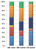
[Infection-surveillance experience at a neurological intensive care unit]
[Infection-surveillance is an important part of the infection control system serving the protection of patients and healthcare workers as well. The continuous surveillance of health care associated infections is among the most important fields of patient safety and quality management. The aim of this study was to evaluate the frequency of the health care associated infections among patients at the neurointensive care unit. Moreover, we aimed to identify specific infectionforms and the most frequently occurring pathogens. We performed the study for a half-year according to the HELICSmethod proposed by the National Center of Epidemiology. In this setup we evaluated the infections and risk factors for infection (instrument-use, antibiotic therapy etc.) among the patients who spent at least 48 hours in the neurointensive care unit. During the six-month period, we observed 16 health care associated mono- and polymicrobial infections out of the 88 cases. Mainly Gram-positive pathogens were identified, but we found multidrug-resistant pathogens as well. Clinically diagnosed pneumonia was the most frequent among the infections. These infections were detected by a relatively low microbiological testing rate, which warns to increase sampling frequency to ensure more accurate data on infections. Infection control based on a comparative standardized infection dataset seems to be one of the most important preventive measures.]
Clinical Neuroscience
OCTOBER 05, 2013
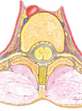
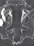
[Split laminotomy and complementary spacer insertion for opening and enlargement of the thoracic spinal canal at infiltrative intramedullary tumor removal]
[Objective - The author main objective was to improve the previously developed technique of split laminotomy and moderate enlargement of the spinal canal with preservation of the majority of posterior structures, and to avoid the complications of the classic autologous bone grafting procedure. Methods - A multilevel spinous process splitting and distracting laminotomy technique with complementary spacer insertion between the laminar parts was developed. We used Poly-Ether-Ether-Ketone (PEEK) cages. This improved method was used in five patients to remove malignant intramedullary tumors at the thoracic level. Results - Adequate surgery of the tumors located intramedullary, and permanent decompression of the spinal canal was achieved in all patients using our new modified procedure. The results have been postoperatively confirmed with MRI and CT. The affected spine was the thoracic in all cases. The numbers of split laminae were three to five. Histological results were as follows: four intramedullary astrocytomas, one ependymoma. The ependymoma was completely, while the astrocytomas were only subtotally removed. In all cases heterologous grafts were inserted between the sides of the distracted laminas, to achieve the enlargement of the spinal canal. The mean duration of the whole surgical procedure was 118 minutes (range 91 to 145 minutes). The average follow-up was 11.2 months, with the range from five to 16 months. Upon postoperative neurological follow-up, no complications were revealed related to the newly developed procedure. The postoperative followup CT scans demonstrated bony healing, with a cage between the osteotomized faces. No compression or dislocation of the spacer was seen. Instability was not detected in any of the patients by flexion or extension lateral radiographs. Conclusion - This modification of the split laminotomy and heterologous grafting method fulfills the requirements of other laminotomy techniques. The split laminotomy is suitable for removing intramedullary tumors, and the posterior stabilizing structures of the spine, as the vertebral laminae and the longitudinal musculature are completely prevented. Due to use of allograft the complications of the classic hip bone grafting procedures are avoided. The spacers, inserted between the osteotomized faces, provided permanent decompression of the spinal canal, and bony healing - throughout the spacer - of the splitted vertebral laminae, without iliac graft complications.]
Clinical Neuroscience
OCTOBER 05, 2013
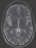
[The diagnosis of herpesencephalitis - a case-based update]
[Herpes simplex virus encephalitis (HSVE) is a rare and lifethreatening infection. The clinical signs are diverse and often misleading regarding the aetiology. However, focal seizure with fewer and typical CT/MRI finding should always raise the possibility of HSVE as early diagnosis and antiviral therapy is crucial. Before the advent of molecular techniques and high-tech imaging histological examination from multiple brain biopsies were often necessary. Although nowadays PCR and other molecular methods may provide an aetiological diagnosis some cases need neuropathological verification. Due to the high IgG seropositivity rate in the population the plasma IgG titer is not diagnostic and elevation of its plasma level requires several weeks. We report the case of a 25-years old male patient who initially presented with epileptic fits. There was no final diagnosis and causal treatment in the district general hospital. The patient was admitted to our institution in comatose state on day 9; the initiated diagnostic tests and therapy could not save the patient who died next day. The autopsy and subsequent neuropathological examination revealed HSVE. We present a flowchart on diagnostic work-up and special techniques to aid diagnosis in suspected viral encephalitis.]
Clinical Neuroscience
OCTOBER 05, 2013
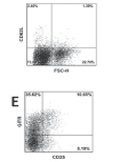
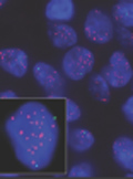
[Characterization of CD4+ and CD8+ Tregs in a Hodgkin’s lymphoma patient presenting with myasthenia-like symptoms]
[The co-occurrence of Hodgkin’s lymphoma (HL) and myasthenia gravis (MG) is a rare phenomenon that is sometimes considered a paraneoplastic manifestation. There are a few documented cases in which myasthenia symptoms manifested only after the surgical removal of the tumor. However, the biological basis of this association is unknown. One hypothesis is that it derives from the infiltration of the residual thymic tissue by the developing tumor. In our case, the myasthenic symptoms led to the HL diagnosis. Our objective was to investigate the T cell phenotype in a HL patient presenting myasthenia-like symptoms. In patients with autoimmune disease, Tregs are usually decreased, but in some diseases, they appear to be increased. It has been speculated that this phenomenon may occur due to a homeostatic attempt by the immune system to control the expansion of auto-reactive effector cells. In the described patient the proportion of lymphoma infiltrating Tregs was high (more than 10% of CD4+ and 1.34% of CD8+ cells), suggesting that Tregs are increased in patients suffering from HL and eventually of myasthenia gravis. Treg involvement in HL is controversial and is currently under investigation. In this context, our data may contribute to a better understanding of the underlying mechanism of the link between HL and autoimmune phenomena.]
Clinical Neuroscience
OCTOBER 05, 2013

Clinical Neuroscience
OCTOBER 05, 2013
Clinical Neuroscience
OCTOBER 05, 2013

Clinical Neuroscience
OCTOBER 05, 2013

Clinical Neuroscience
OCTOBER 05, 2013

1.
Clinical Neuroscience
[Headache registry in Szeged: Experiences regarding to migraine patients]
21. MAY
2.
Clinical Neuroscience
[The new target population of stroke awareness campaign: Kindergarten students ]
21. MAY
3.
Clinical Neuroscience
Is there any difference in mortality rates of atrial fibrillation detected before or after ischemic stroke?
27. NOV
4.
Clinical Neuroscience
Factors influencing the level of stigma in Parkinson’s disease in western Turkey
27. SEP
5.
Clinical Neuroscience
[The effects of demographic and clinical factors on the severity of poststroke aphasia]
18. JUL
1.
2.
3.
4.
5.