The eLitMed.hu medical portal uses computer cookies for convenient operation. Detailed information can be found in the Cookie-policy.
Clinical Neuroscience - 2012;65(07-08)
Content
Clinical Neuroscience
JULY 30, 2012


Clinical Neuroscience
JULY 30, 2012

[Neurophobia]
[Neurophobia is the fear of neurological diseases. Its main symptom is that medical students and young doctors are not able to utilize their basic neurological knowledge at the bedside. According to statistics, every second student suffers from neurophobia. This attitude could explain why in the last two decades less and less young doctors wanted to become neurologist. Medical students complain that they receive no instructions, and are afraid of loosing their interest and of facing the failure of their competency. The hardship of neurology was explained by the insufficient knowledge of anatomy and the infrequent encounter with patients. Even general practitioners have anxiety about neurological patients. The loss of interest in neurosciences seems to associate with insensitivity of human-centered culture and corruption of empathic thinking. The burnout syndrome of medical doctors and students can be explained by stress, loss of respect, permanent competition, independency that interferes with responsibility, stiff hierarchy of medical society, fear of diagnostic failures and of economical difficulties. The scores of depression in female students were higher than in male. The idea of the “good neurologist” has been changed. The business oriented care, the shortage of time, and the financial restrictions corroded the conventional practice and ceased the vocational idealism. At present, personal teaching is going to transform into impersonal multimedia learning. Because of the drastic change of values, the age of inner-oriented professionals has terminated also in the medicine. Medical doctors follow even less the traditional troll of professional behavior, but according the social demands, they choose their specialization for subsistence. The highly esteemed social status of neurologists and psychiatrists is going to sink in Europe. To reduce neurophobia it would be desirable 1. to introduce neurology training in the early years of medical school; 2. to teach neurology in all semesters, 3. to assure the effective teaching of neuro-anatomy and physiology, 4. to organize more one-to-one teacher-student communication. In the United States, residents participate in teaching during their residency training. To master neurology dedicated teachers are necessary whom neurology residents ought to meet personally with optimal frequency. However, these requirements seem to fail because of the chiefly technical characters of the actual reforms.]
Clinical Neuroscience
JULY 30, 2012
[Diagnosis and therapy of mitochondrial diseases]
[Mitochondrial diseases are a significant part of neuromuscular diseases. Majority of them is multisystemic disorder. The diagnosis can be established in more and more cases. Beyond the routine neurological examination imaging methods (MRI and MR-spectroscopy) and electrophysiology (EMG, ENG, EEG, evoked potential tests) might be helpful in setting the diagnosis. Raised blood lactate level supports the diagnosis. Muscle biopsy demonstrates mitochondrial abnormalities in the majority of cases. The positivity of genetic tests is low, because the amount of mitochondrial DNA alterations is different in tissues. Therefore other tissue than blood (mainly muscle) is necessary for genetic tests. The other reason is that the respiratory chain is under double -mitochondrial and nuclear - genetic control, and testing the nuclear genes are available only in selected laboratories. The treatment is limited, mainly symptomatic.]
Clinical Neuroscience
JULY 30, 2012

[The absence of the common LRRK2 G2019S mutation in 120 young onset Hungarian Parkinon’s disease patients]
[Parkinson’s disease is a promising target of applying personalized medicine. For this purpose it is crucial to reveal the genetic and environmental factors, which contribute to the disease, also to collect epidemiologic data and to preserve the patients samples and data in a proper biobank. In our investigation we examined the prevalence of the most frequent Parkinson’s disease causing LRRK2 G2019S mutation in a Hungarian Parkinson-patient group. From 120 patients, we haven’t detected this substitution in anyone. Our investigation suggest that the mutation LRRK2 G2019S may be a rare cause of Parkinson disease in the Hungarian population.]
Clinical Neuroscience
JULY 30, 2012

[Results of intrathecal baclofen therapy on spasticity in patients with brain injury]
[Objectives - To evaluate the results of intrathecal baclofen (ITB) therapy on the spasticity in patients with brain injury. Method - Retrospective study in Brain Injury Rehabilitation Unit between January 2001 and December 2010. Results - During the last ten years, in our unit 13 patients were involved into ITB therapy on severe spasticity, after brain injury, while more than 100 Baclofen pumps were implantated in Hungary with Hungary with coordination of the Multidisciplinary Team. ITB therapy was indicated in severe spasticity developed after seven cases of traumatic brain injuries, five cases of strokes and one case of anoxic brain injury. The mean age of patients was 26 years (18- 52). At the time of pump implantation three patients were in vegetative state. The shortest period elapsed between the brain injury and pump implantation was three months and the longest period was nine years, mean 15 months. Baclofen pump had to be changed in six cases after six years, and was removed in three cases due to decreasing spasticity. Catheter revision was performed in two cases due to flow problem. We had no complication in association with ITB therapy. Conclusions - Intrathecal baclofen therapy seems to be an effective and safe treatment in patients with severe spasticity of cerebral origin. We suggest team (neurosurgeon and rehabilitation professionals) decision in a spasticity center before involving the patient into ITB therapy, and follow up in the rehabilitation unit. The severity of spasticity as a consequence of brain injury can change during years and it is necessery to follow it with dosage and dynamics of baclofen therapy. Baclofen pump removal is suggested if the ITB therapy is further not reasonable.]
Clinical Neuroscience
JULY 30, 2012

Clinical Neuroscience
JULY 30, 2012

[Treatment of dystonia by deep brain stimulation: a summary of 40 cases]
[Background - Bilateral pallidal deep brain stimulation (DBS) is an established treatment option for primary generalized and segmental dystonia. In the present study we evaluated the results of our dystonia patients treated by DBS. Methods - The surgical results of forty consecutive dystonia patients underwent DBS implantation were analyzed (age: 43.7±17.7 years; sex: 22 men; etiology: 24 primary and 16 secondary dystonia; topography: 24 generalized, 12 segmental and four hemidystonia; disease duration: 16.1±9.3 years). Severity of dystonia measured by Burke-Fahn-Marsden Dystonia Rating Scale (BFMDRS) and health-related quality of life measured by EQ-5D scale were obtained preoperatively and compared to the scores obtained at postoperative six months and subsequent yearly follow-ups. The average follow-up lasted 2.5 years (median, 0.5-8 years). In all cases the BFMDRS scores were re-evaluated by a rater blinded to the treatment. Treatment responsiveness was defined as an at least 25% improvement on the BFMDRS scores. Non-parametric Mann-Whitney, McNemar and Kruskal-Wallis tests were applied to test statistical significance. Results - Severity of dystonia improved from 31 to 10 points (median, 68% improvement, p<0.01) in the primary dystonia group, whereas in secondary dystonia these changes were statistically insignificant (improvement from 40 to 31.5 points, 21.2%, p>0.05). However, the health-related quality of life significantly improved in both groups (primary dystonia: 0.378 vs. 0.788 and secondary dystonia: 0.110 vs. 0.388, p<0.01). Significantly more patients in the primary dystonia group responded to DBS treatment than those in the secondary dystonia group (83.3% vs. 37.5%, p<0.01). Conclusion - Our results are in accordance with previously published international findings demonstrating that DBS is a highly effective and long-lasting treatment option for primary dystonia. DBS is considerably less efficient in secondary dystonia; however, it still has a high impact on the quality of life presumably due to its pain-relieving effect.]
Clinical Neuroscience
JULY 30, 2012

[Are oppressive dreams indicators in bereavement?]
[Objectives - It is widely believed that oppressive dreams are frequent in bereavement - despite the lack of scientific investigations of the subject. The aims of our study were the analysis of dream quality as well as the correlates of oppressive dreams in bereavement. Method - Participants with (N=473) and without bereavement were compared upon the database of a national representative study (Hungarostudy Epidemiological Panel Survey 2006, N=4329). Dream contents were assessed with the Dream Quality Questionnaire (DQQ). Depressive symptoms (BDI-S) and the presence anxiety were also investigated. Results - Oppressive dreams occurred significantly higher frequency in the first year of bereavement (men: F=17.525, p<0.001, women: F=8.291, p=0.004). Oppressive dreams were significantly associated with anxiety (F=37.089, p<0.001) and with depressive symptoms (F=50.562, p<0.001). Discussion - Oppressive dreams are significantly more frequent in the first year of bereavement, and may act as indicators of bereavement-linked mental health consequences like depression and anxiety. These are often masked by the symptoms of grief and therefore remain untreated. Our preliminary results could be a starting point for the development of further research aiming to clarify the relationship amongst dream contents, anxiety, and depression in bereavement.]
Clinical Neuroscience
JULY 30, 2012
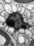
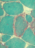
[Selective ultrastructural vulnerability in the cuprizone-induced experimental demyelination]
[Background and purpose - It has been reported that multiple sclerosis has four different neuropathological subtypes, and two of them (type III and IV) are characterized by primary oligodendrocyte loss. However, the exact pathomechanism that lead to oligodendrocyte apoptosis in human demyelinating diseases is still elusive. The copper chelator cuprizone induces primary oligodendrocyte apoptosis and consequent demyelination in well defined areas of the mouse brain. Nevertheless, the precise subcellular events that result in oligodendrocyte cell death in the cuprizone model are still unknown. We aimed to study the ultrastructural alterations that might induce oligodendrocyte apoptosis in the cuprizone experimental demyelination model. Methods - C57BL/6 mice were given cuprizone for two, 21 and 35 days to induce demyelination to investigate early pathological events, and different stages of demyelination. In addition, mice were given cuprizone for 35 days and were allowed to recover for two or 14 days to study early and late remyelination. After the cuprizone treatment, mice were sacrificed and the corpus callosum, the superior cerebellar peduncle, the optic nerve and the sciatic nerve were studied by electron microscopy. Results - The ultrastructural analysis revealed that cuprizone induced oligodendrocyte apoptosis is accompanied by the formation of giant mitochondria in the affected cells in the corpus callosum and in the superior cerebellar peduncle. Apoptosis of the myelin producing cells was present through the whole cuprizone challenge. Severe demyelination occurred after three weeks of cuprizone administration associated with massive macrophage infiltration and astrocytosis of the demyelinated areas. Axons and neurons remained unaffected. Conclusion - The formation of giant mitochondria in myelin producing oligodendrocytes is the first pathological sign in the cuprizone experimental demyelination. Mitochondrium pathology in the cuprizone challenge might serve as a useful model to study the pathomechanism of multiple sclerosis subtypes (III and IV) characterized by primary oligodendrocyte degeneration.]
Clinical Neuroscience
JULY 30, 2012
[Endoscopic, posterior transseptal pituitary surgery - Learning curve of the surgical technique and equipment in 61 operations]
[Introduction - The removal of hypophyseal tumor by transsphenoidal pituitary surgery using microsurgical instruments was first performed over 100 years ago. Operating techniques for this surgery are constantly being renewed, first by using a microscope and later on with the use of an endoscop. The authors provide an overview of the minimal invasive posterior transseptal-transsphenoidal aproach with the combined utilization of classical techniques with the assistance of the endoscop. Method - Sixty-one patients (33 female, 28 male, 21-84 yrs) were treated for sellar region tumor resection using an endonasal transsphenoidal aproach with the help of an endoscop. Follow ups were performed within 2-21 months. Results - Total tumor resection was successful in 91.8%, and partial resection in 8.2% of the patients. The rate of complications using the endoscop method was not higher compared to that of the classical microscopic method. There was no major bleeding in any of the cases. Adverse events such as minor epistaxis occurred in 4.9%, transitional diabetes insipidus in 6.5%, inraoperative CSF leak in 16.67%, postoperative CSF leak in 11.5% and meningitis in 8.2% of the patients. After the operation the pathological hormonal production stoped in all patients except in two patients who were acromegalic. However their GH level normalized and they did not require further treatment, the IGF-1 still remained high. Conclusion - The success of the surgical treatment is based on both, the proficient pre- and postoperative endocrinological care, and the minimal invasive surgical technique. The endoscop was used partially or continuously during the operation for better visualization of the operation field in multiple angles (30°, 45°). It was useful in differentiating between normal and tumorous glandular tissue, and also offered an enhanced view of the intrasellar (via hydroscopy) and parasellar region. Moreover the endoscopic method is able to decrease the operating time, reduce blood loss. In different stages of the surgery, depending on the anatomical and pathological situation, switching back and forth from microscope to endoscop technique, gives us the benefit of a clearer view in each situation.]
Clinical Neuroscience
JULY 30, 2012

Clinical Neuroscience
JULY 30, 2012

Clinical Neuroscience
JULY 30, 2012

1.
Clinical Neuroscience
[Headache registry in Szeged: Experiences regarding to migraine patients]
21. MAY
2.
Clinical Neuroscience
[The new target population of stroke awareness campaign: Kindergarten students ]
21. MAY
3.
Clinical Neuroscience
Is there any difference in mortality rates of atrial fibrillation detected before or after ischemic stroke?
27. NOV
4.
Clinical Neuroscience
Factors influencing the level of stigma in Parkinson’s disease in western Turkey
27. SEP
5.
Clinical Neuroscience
[The effects of demographic and clinical factors on the severity of poststroke aphasia]
18. JUL
1.
2.
3.
4.
5.