The eLitMed.hu medical portal uses computer cookies for convenient operation. Detailed information can be found in the Cookie-policy.
Hypertension and nephrology - 2012;16(06)
Content
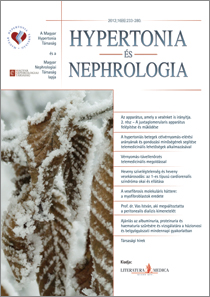
[The apparatus which controls our kidney too. Part 2 - Structure and function of the juxtaglomerular apparatus]
[The juxtaglomerular apparatus is comprised of the macula densa (a specialregion of the distal tubule), the renin producing granular or epithelioid-cells of the afferent arteriole, the extraglomerular mesengial cells, and the efferent arteriole’s section bordering this region. Somewhat more general definitions also exist. Recently, distinctive morphological and functional associations have been identified between the components of the JGA and some common mediators (e.g. adenosine, angiotensin, NO, prostaglandins, etc.). Current data suggest that each cell of the macula densa also contain few cilia that may have a role in sensing fluid flow. The distal section of the afferent arteriole (possessing the same structure as the glomerular capillaries) is covered by fenestrated endothelium. Trace dose of ferritin particles can pass through the afferent arteriole’s fenestrae into the interstitium of the JGA due to the considerable hydrostatic pressure gradient. The parietal lamina of Bowman’s capsule, which covers the renin granulated cells of the afferent arteriole behaves much like the visceral lamina in that the epithelial cells of the parietal lamina exhibit foot processes and filtration slits. The urinary space is regularly bulging into the extraglomerular mesangium. Therefore, the notion has been refuted that the JGA, which contains neither blood nor lymph capillaries, is a closed system engaging in only slow fluid exchange. Furthermore it is affirmed that the afferent arteriole consists of two morphologically and functionally disparate segments, the ratio of which is considerably modified by the activity of the renin-angiotensin system.]
[The improvement of the rate of reaching the target blood pressure and the quality of care of hypertensive patients with applying telemedicine facilities]
[The authors summarize the facilities of the telemedicine and telemonitoring system. The methods of telemedicine and their combination with home blood pressure measurement or the help of a nurse or pharmacist are reviewed. In the light of the latest results the authors are led to the conclusion that the intensive spread of using the facilities of telemedicine is necessary in the present-day Hungarian healthcare system. At the same time it is also determined that the methodical and technical potential is not enough to further enhance the efficiency in itself. The personal contact and the possibility of interactive monitoring from distance are definitely crucial for the continuous maintenance of the reached target blood pressure in patients suffering from hypertension and also for the augmentation of patient adherence regarding applying medicine.]
[Blood pressure self-measurement with telemonitoring technology]
[Authors present the guidelines, indications and utility value of home selfmeasurements of blood pressure. They report the results of the most important clinical studies. They analyze the methodology of the measurements within telemedicinal solutions and describe the consultative scopes associated with the measurement methods already applied in clinical practice. Their own telemonitoring system - called Medistance - is then presented. They have created three modules for the long term registration of blood pressure in hypertensive patients: 1. an individual module for the hypertensive patients, the elderly, the family, for patients with high cardiovascular risk and for the physicians. 2. a module for the pharmaceutical care, 3. a module for the communities (social homes, club for the elderly, etc.). The Medistance system is functioning for two years in our count]
[Acute heart failure and acute renal injury: pathophysiology and management of cardiorenal syndrome type 1]
[The functional connection between heart and kidney is well known. Several types of this relationship have been recently characterized as cardiorenal syndromes. The relevance of this relationship in clinical practice is supported by the fact, that the consequences of the primary dysfunction are profoundly influenced by the magnitude and the treatment possibilities of the secondary dysfunction. Moreover, the administered therapy for heart failure can deteriorate renal hemodinamics, or side effects of the treatment can lead to the worsening of the clinical picture. Loop diuretics decrease venous congestion, but also induce neurohormonal activation and a decrease in glomerular filtration rate. The body of positive evidence with the use of mineralocorticoid receptor antagonists in acute settings is limited. Inotropic agents on the one hand improve hemodinamics, on the other hand increase the danger of arrhythmia and mortality (levosimendan seems to be an exception). Aquaretics decrease symptoms without influencing mortality. The natriuretic peptide neseritid improved clinical symptoms, but did not improve endpoints in clinical trials. Vasodilators improve hemodinamics, but their usefulness is limited because of their profound hypotensive effect. The effectiveness and benefits of ultrafiltration has to be tested in more clinical trials. Because of such treatment difficulties the management of these patients is a complex task that needs the involvement of intensive therapeutic specialists, nephrologists and cardiologists. This review focuses on the pathophysiology and possible management of the patients with acute heart failure with acute kidney injury, called type 1 cardiorenal syndrome from the cardiologist’s point of view.]
[Molecular mechanisms leading to renal fibrosis: the origin of myofibroblasts]
[There are about a quarter of million patients who need chronic renal replacement therapy in Europe, and the estimated number of patients with chronic kidney disease is about tenfold higher. Interestingly, regardless of the initiating cause the mechanism of fibrosis is similar to each other in the different chronic kidney diseases. In general, the damaged glomerular or tubular cells release danger signals and produce chemotactic stimuli, which trigger the rapid recruitment of leukocytes. The infiltrating immune cells and the damaged renal cells then produce high levels of proinflammatory cytokines, growth factors, chemokines and adhesion molecules which contribute to glomerular/tubular injury, accumulation of further leukocytes and myofibroblasts, which are the effector cells of renal fibrosis. However the origin of myofibroblasts is still controversial. Recent hypotheses suggest that they are originated from different renal cells, such as epithelial and endothelial cells, pericytes or bone marrow derived fibrocytes. The myofibroblasts thus generated serve as key cellular mediators of renal fibrosis. Myofibroblasts have migratory capacity, are resistant to apoptosis, produce several growth factors and cytokines and according to our present knowledge these cells are the main source of collagen-I and -III rich extracellular matrix in the fibrous tissue. Organ fibrosis is characterized with excessive deposition of extracellular matrix leading to glomerular sclerosis and renal tubulointerstitial fibrosis. The excessive deposition of fibrous tissue replaces healthy kidney tissue; nephrons disappear and kidney function declines gradually. In this article the knowledge is summarized on the molecular changes leading to the generation of renal myofibroblasts.]
1.
Clinical Neuroscience
Is there any difference in mortality rates of atrial fibrillation detected before or after ischemic stroke?2.
Clinical Neuroscience
Factors influencing the level of stigma in Parkinson’s disease in western Turkey3.
Clinical Neuroscience
Neuropathic pain and mood disorders in earthquake survivors with peripheral nerve injuries4.
Journal of Nursing Theory and Practice
[Correlations of Sarcopenia, Frailty, Falls and Social Isolation – A Literature Review in the Light of Swedish Statistics]5.
Clinical Neuroscience
[Comparison of pain intensity measurements among patients with low-back pain]1.
Clinical Neuroscience Proceedings
[A Magyar Stroke Társaság XVIII. Kongresszusa és a Magyar Neuroszonológiai Társaság XV. Konferenciája. Absztraktfüzet]2.
3.
Journal of Nursing Theory and Practice
[A selection of the entries submitted to the literary contest "Honorable mission: the joys and challenges of our profession" ]4.
Journal of Nursing Theory and Practice
[End of Life and Palliative Care of Newborns in the Nursing Context]5.
Journal of Nursing Theory and Practice
[Aspects of Occupational Health Nursing for Incurable Patients ]


