Clinical Neuroscience

Electrophysiological investigation for autonomic dysfunction in patients with myasthenia gravis: A prospective study

NALBANTOGLU Mecbure1, AKALIN Ali Mehmet2, GUNDUZ Aysegul3, KIZILTAN Meral3

JANUARY 30, 2021

Clinical Neuroscience - 2021;74(01-02)

DOI: https://doi.org/10.18071/isz.74.0033

Journal Article

Szöveg nagyítása:

-
+

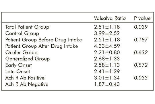
Myasthenia gravis (MG) is an autoimmune disorder of neuromuscular transmission. Autonomic dysfunction is not a commonly known association with MG. We conducted this study to evaluate autonomic functions in MG & subgroups and to investigate the effects of acetylcholinesterase inhibitors. This study comprised 30 autoimmune MG patients and 30 healthy volunteers. Autonomic tests including sympathetic skin response (SSR) and R-R interval variation analysis (RRIV) was carried out. The tests were performed two times for patients who were under acetylcholinesterase inhibitors during the current assessment. The RRIV rise during hyperventilation was better (p=0.006) and Valsalva ratio (p=0.039) was lower in control group. The SSR amplitudes were lower thereafter drug intake (p=0.030). As much as time went by after drug administration prolonged SSR latencies were obtained (p=0.043).Valsalva ratio was lower in the AchR antibody negative group (p=0.033). The findings showed that both ocular/generalized MG patients have a subclinical parasympathetic abnormality prominent in the AchR antibody negative group and pyridostigmine has a peripheral sympathetic cholinergic noncumulative effect.

AFFILIATIONS

  1. Demiroglu Bilim University Medical Faculty, Neurology Department, Istanbul, Turkey
  2. Memorial Bahcelievler Hospital, Neurology Department, Istanbul, Turkey
  3. Istanbul University Cerrahpasa Medical Faculty, Neurology Department, Istanbul, Turkey

COMMENTS

0 comments

Further articles in this publication

Clinical Neuroscience

Cases of inborn errors of metabolism diagnosed in children with autism

CAKAR Emel Nafiye, YILMAZBAS Pınar

Autism spectrum disorder is a neurodevelopmental disorder with a heterogeneous presentation, the etiology of which is not clearly elucidated. In recent years, comorbidity has become more evident with the increase in the frequency of autism and diagnostic possibilities of inborn errors of metabolism. One hundred and seventy-nine patients with diagnosis of autism spectrum disorder who presented to the Pediatric Metabolism outpatient clinic between 01/September/2018-29/February/2020 constituted the study population. The personal information, routine and specific metabolic tests of the patients were analyzed retrospectively. Out of the 3261 patients who presented to our outpatient clinic, 179 (5.48%) were diagnosed with autism spectrum disorder and were included in the study. As a result of specific metabolic examinations performed, 6 (3.3%) patients were diagnosed with inborn errors of metabolism. Two of our patients were diagnosed with classical phenylketonuria, two with classical homocystinuria, one with mucopolysaccharidosis type 3D (Sanfilippo syndrome) and one with 3-methylchrotonyl Co-A carboxylase deficiency. Inborn errors of metabolism may rarely present with autism spectrum disorder symptoms. Careful evaluation of the history, physical examination and additional findings in patients diagnosed with autism spectrum disorder will guide the clinician in the decision-making process and chose the appropriate specific metabolic investigation. An underlying inborn errors of metabolism may be a treatable cause of autism.

Clinical Neuroscience

Evaluation of the effectiveness of transforaminal epidural steroid injection in far lateral lumbar disc herniations

EVRAN Sevket, KATAR Salim

Far lateral lumbar disc herniations (FLDH) consist approximately 0.7-12% of all lumbar disc herniations. Compared to the more common central and paramedian lumbar disc herniations, they cause more severe and persistent radicular pain due to direct compression of the nerve root and dorsal root ganglion. In patients who do not respond to conservative treatments such as medical treatment and physical therapy, and have not developed neurological deficits, it is difficult to decide on surgical treatment because of the nerve root damage and spinal instability risk due to disruption of facet joint integrity. In this study, we aimed to evaluate the effect of transforaminal epidural steroid injection (TFESI) on the improvement of both pain control and functional capacity in patients with FLDH. A total of 37 patients who had radicular pain caused by far lateral disc herniation which is visible in their lumbar magnetic resonance imaging (MRI) scan, had no neurological deficit and did not respond to conservative treatment, were included the study. TFESI was applied to patients by preganglionic approach. Pre-treatment Visual Analogue Scale (VAS) and Oswestry Disability Index (ODI) scores of the patients were compared with the 3rd week, 3rd month and 6th month scores after the procedure. The mean initial VAS score was 8.63 ± 0.55, while it was 3.84 ± 1.66, 5.09 ± 0.85, 4.56 ± 1.66 at the 3rd week, 3rd month and 6th month controls, respectively. This decrease in the VAS score was found statistically significant (p = 0.001). ODI score with baseline mean value of 52.38 ± 6.84 was found to be 18.56 ± 4.95 at the 3rd week, 37.41 ± 14.1 at the 3rd month and 34.88 ± 14.33 at the 6th month. This downtrend of pa­tient’s ODI scores was found statistically significant (p = 0.001). This study has demonstrated that TFESI is an effective method for gaining increased functional capacity and pain control in the treatment of patients who are not suitable for surgical treatment with radicular complaints due to far lateral lumbar disc hernia.

Clinical Neuroscience

Neuroscience highlights: The mirror inside our brain

KRABÓTH Zoltán, KÁLMÁN Bernadette

Over the second half of the 19th century, numerous theories arose concerning mechanisms involved in understanding of action, imitative learning, language development and theory of mind. These explorations gained new momentum with the discovery of the so called “mirror neurons”. Rizzolatti’s work inspired large groups of scientists seeking explanation in a new and hitherto unexplored area of how we perceive and understand the actions and intentions of others, how we learn through imitation to help our own survival, and what mechanisms have helped us to develop a unique human trait, language. Numerous studies have addressed these questions over the years, gathering information about mirror neurons themselves, their subtypes, the different brain areas involved in the mirror neuron system, their role in the above mentioned mechanisms, and the varying consequences of their dysfunction in human life. In this short review, we summarize the most important theories and discoveries that argue for the existence of the mirror neuron system, and its essential function in normal human life or some pathological conditions.

Clinical Neuroscience

Matrix metalloproteinases and their tissue inhibitors in relapsing remitting multiple sclerosis: Possible markers and treatment agents

SANLI Arzu, OZTURK Musa, SOYSAL Aysun, DOVENTAS Yasemin, BASOGLU Fulya, GOZUBATIK-CELIK R. Gokcen, BAYBAS Sevim

Matrix metalloproteinases (MMPs), which are synthesized by many cell groups and responsible for the destruction of matrix proteins, and endogen tissue inhibitors of MMPs (TIMPs) have a role in the pathogenesis of Multiple Sclerosis (MS) by affecting the blood-brain barrier. We aimed to investigate the role of MMPs and TIMPs in the immunopathogenesis and in the course of multiple sclerosis (MS). We enrolled 25 relapsing remitting MS patients, who had a definite MS diagnosis according to McDonald criteria and 25 healthy subjects similar for age and gender as control group. MMP-9- and TIMP-1 levels were measured twice in patient group (one time during an attack and one in remission) and once in healthy subjects. MMP-9- and TIMP-levels of patients during attack and remission period and MMP-9/TIMP-1 ratio were found significantly higher than in the control subjects. In patient group MMP-9 and TIMP-1 levels and MMP-9/TIMP-1 ratio during attacks were not significantly different than during remission period. However, when subdivided according to their number of attacks, patients with 2 attacks had significantly higher levels during attack period comparing to remission period (p<0.05); in case of patients with more than 2 attacks did not have a statistically significant difference in attack and remission periods. Matrix metalloproteinases are important actors in MS immunopathogenesis, particularly in the early period and inhibitor agents for these enzymes can be used as a treatment option.

Clinical Neuroscience

Retinal morphological changes during the two years of follow-up in Parkinson’s disease

ATUM Mahmut, DEMIRYÜREK Enes Bekir

The study aims to investigate the relationship between the progression of idiopathic Parkinson’s disease (IPD) and retinal morphology. The study was carried out with 23 patients diagnosed with early-stage IPD (phases 1 and 2 of the Hoehn and Yahr scale) and 30 age-matched healthy controls. All patients were followed up at least two years, with 6-month intervals (initial, 6th month, 12th month, 18th month, and 24th month), and detailed neurological and ophthalmic examinations were performed at each follow-up. Unified Parkinson’s Disease Rating Scale part III (UPDRS Part III) scores, Hoehn and Yahr (H&Y) scores, best-corrected visual acuity (BCVA), intraocular pressure (IOP) measurement, central macular thickness (CMT) and retinal nerve fiber layer (RNFL) thickness were analyzed at each visit. The average age of the IPD and control groups was 43.96 ± 4.88 years, 44.53 ± 0.83 years, respectively. The mean duration of the disease in the IPD group was 7.48 ± 5.10 months at the start of the study (range 0-16). There was no statistically significant difference in BCVA and IOP values between the two groups during the two-year follow-up period (p> 0.05, p> 0.05, respectively). Average and superior quadrant RNFL thicknesses were statistically different between the two groups at 24 months and there was no significant difference between other visits (p=0.025, p=0.034, p> 0.05, respectively). There was no statistically significant difference in CMT between the two groups during the follow-up period (p> 0.05). Average and superior quadrant RNFL thicknesses were significantly thinning with the progression of IPD.

All articles in the issue

Related contents

Clinical Neuroscience

Effects of rituximab on prognosis in myasthenia gravis: A single-center experience from Turkey

GÖL Fatih Mehmet , KARA Fatma , BOZ Merve , MUTLU Abdüssamet , KARAKULLUKÇU Serdar , BOZ Cavit

Management of treatment-resistant patients with myasthenia gravis (MG) remains an important issue. This study aimed to evaluate the effects of rituximab (RTX) treatment on the prognosis of patients with acetylcholine receptor autoantibody-positive (AChR-Ab+), muscle-specific kinase autoantibody-positive (MuSK-Ab+), or seronegative or double seropositive MG.

Nineteen patients treated with RTX between 2015 and 2020 were included in this study. Demographic and clinical characteristics, prognosis, and prognostic predictors of MG were evaluated retrospectively. The Myas­thenia Gravis Foundation of America Post-Inter­vention Status (MGFA-PIS) before RTX treatment (pre-RTX) and after RTX treatment (post-RTX) were recorded.

A total of 10 patients (52.6%) were AchR Ab+, 6 patients (31.6%) were MuSK Ab+, 1 patient (5.3%) was seronegative, and 2 patients (10.5%) were double seropositive. Steroid dose was pre-RTX 38.9±5.7 (25-45), it was post-RTX 10.5±10.3 (0-30) (p<0.001). Post-RTX steroid treatment was discontinued in 6 of 19 patients (p=0.041). Only three patients received intravenous immunoglobulin at the post-RTX follow-up (p<0.001). In post-RTX 12th month, the MGFA-PIS score was as minimally manifestation or better in 9 patients (47.3%) and improved or was better in 18 patients (94.7%) (p-value 0.004; <0.001, respectively).

The improvement in MGFA-PIS scores post-RTX was similar in MuSK-Ab+ and AChR-Ab+ patients. The data are insufficient in seronegative and double seropositive patients and RTX must be considered in the treatment of suitable patients with MuSK-Ab+ and AChR-Ab+ refractory MG.

Clinical Neuroscience

Pembrolizumab-induced peripheral nervous system damage: A combination of myositis/ myasthenia overlap syndrome and motor axonal polyneuropathy

ARMAN ÇAKAR, IBRAHIM KAMACI, ELIF Kocasoy ORHAN, HACER DURMUŞ, YEŞIM PARMAN

Immune-checkpoint inhibitors (ICI) are effective drugs in cancer treatment that block immune checkpoints and stimulate an attack on cancer cells. However, various side effects were reported with ICIs. Peripheral nervous system (PNS) side effects are three times more frequent than those in the central nervous system. A 63-year-old male patient was admitted to our department with a 10-day history of dyspnea, diplopia, and generalized weakness.

Clinical Neuroscience

Myasthenia gravis, Guillain-Barré syndrome, or both?

ERDOGAN Cagdas, TEKIN Selma, ÜNLÜTÜRK Zeynep, GEDIK Korkut Derya

Myasthenia gravis (MG) and Guillain-Barré syndrome (GBS) are autoimmune disorders that may cause weakness in the extremities. The coexistence of MG and GBS in the same patient has rarely been reported previously. A 52-year-old male presenting with ptosis of the left eye that worsened with fatigue, especially toward evening, was evaluated in our outpatient department. His acetylcholine receptor antibody results were positive, supporting the diagnosis of MG. His medical history revealed a post-infectious acute onset of weakness in four extremities, difficulty in swallowing and respiratory failure, which was compatible with a myasthenic crisis; however, his nerve conduction studies and albuminocytologic dissociation at the time were compatible with GBS. With this case report, we aimed to mention this rare coincidental state, discuss possible diagnoses and review all other similar cases in the literature with their main features.

Clinical Neuroscience

Autonomic nervous system may be affected after carpal tunnel syndrome surgery: A possible mechanism for persistence of symptoms after surgery

ONDER Burcu, KELES Yavuz Betul

After carpal tunnel surgery, some patients report complaints such as edema, pain, and numbness. Purpose – The aim of this study was to evaluate autonomic nervous system function in patients with a history of carpal tunnel surgery using sympathetic skin response (SSR). Thirty three patients (55 ±10 years old) with a history of unilateral operation for carpal tunnel syndrome were included in the study. The SSR test was performed for both hands. Both upper extremities median and ulnar nerve conduction results were recorded. A reduced amplitude (p=0.006) and delayed latency (p<0.0001) were detected in the SSR test on the operated side compared to contralateral side. There was no correlation between SSR and carpal tunnel syndrome severity. Although complex regional pain syndrome does not develop in patients after carpal tunnel surgery, some of the complaints may be caused by effects on the autonomic nervous system.

Clinical Neuroscience

[Myasthenia in a patient with sarcoidosis and schizophrenia (in English language)]

RÓZSA Csilla, KIS Gábor, KOMOLY Sámuel

[A 44-year-old male patient was hospitalised with paranoid schizophrenia in 1985. Depot neuroleptic treatment was started which successfully prevented further psychotic relapses for the next ten years. His myasthenia gravis started with bulbar signs in 1997 and the symptoms soon became generalized. The diagnosis of myasthenia gravis was confirmed by electromyography, by positive anticholinesterase test and by the detection of anti-acetylcholine receptor antibodies in the serum. Mediastinal CT examination showed enlarged hilar lymph nodes on the left but no thymic pathology was observed. Mediastinoscopy was performed and biopsies were obtained from the affected nodes. Histology revealed sarcoidosis. The patient suffered respiratory crisis following the thoracic intervention (in September 1998). Combined oral corticosteroid (64 mg methylprednisolone/e.o.d.) and azathioprine (150 mg/day) treatment regimen was initiated and complete remission took place in both the myasthenic symptoms and the sarcoidosis. The follow-up CT scans showed no mediastinal pathology (January 2000). During steroid treatment a transient psychotic relapse occured which was successfully managed by supplemental haloperidol medication added to his regular depot neuroleptics. The patient currently takes 150 mg/day azathioprine and receives 40 mg/month flupentixol depot im. His physical and mental status are stable and he has been completely symptome free in the last 24 months. The association of myasthenia gravis and sarcoidosis is very rare. To our best knowledge no case has been reported of a patient suffering from myasthenia gravis, sarcoidosis, and schizophrenia at the same time.]