The eLitMed.hu medical portal uses computer cookies for convenient operation. Detailed information can be found in the Cookie-policy.
Clinical Neuroscience - 2018;71(03-04)
Content
Clinical Neuroscience
MARCH 30, 2018

[The role of MRI in measuring the effectivity of disease modifying treatments I]
[MRI has a significant role in the diagnosis of multiple sclerosis. The newer and newer treatment options of the disease make it necessary to monitor the effectiveness of the therapy. Besides the clinical signs (clinical relapses and progression), the different MRI parameters can also reflect the disease activity. In our current article we summarize those MRI markers, which best predict the long-term disability, based on the international standards.]


Clinical Neuroscience
MARCH 30, 2018
[The role of MRI in measuring the effectivity of disease modifying treatments II]
[The paraclinical examinations, principally the MRI have an increasing significance in the diagnosis of multiple sclerosis. However, MRI markers also have a prominent role in monitoring of the disease-course and activity, and also in the planning of possible therapeutic changes. In accordance with previously published international guidelines, in this article we propose a protocol for the monitoring the treatment efficacy in multiple sclerosis. This could be the basis of a consensus based guideline to be implemented in Hungary.]
Clinical Neuroscience
MARCH 30, 2018
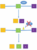
[Nusinersen in the treatment of spinal muscular atrophy]
[Until recently, the diagnosis of spinal muscular atrophy (SMA) has been associated with severe life-long motor disability in adults and with early death in infants. The new experimental therapeutic approaches of the last few years have become more and more promising, while nusinersen was approved for the treatment of SMA in December 2016 in the USA, and in May 2017 in Hungary. Our paper presents mechanisms and clinical benefits of this new medication, and highlights some of the other therapeutic strategies still in experimental stages.]
Clinical Neuroscience
MARCH 30, 2018
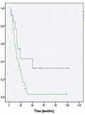
Long-term follow-up results of concomitant chemoradiotherapy followed by adjuvant temozolomide therapy for glioblastoma multiforme patients. The importance of MRI information in survival: Single-center experience
Introduction - Glioblastoma multiforme (GBM) is the most common malignant primary anomaly of central nervous system. The GBM infiltrates the nearly sturctures from the initial tumor and its metastatic attribution is well known. The aim of our single-centered retrospective study was to introduce the importance of postoperative medical imaging confirmation of total tumor resection for patient with GBM combined concomitant and adjuvant chemoradiotherapy on a 10 year long patient follow up. Methods - From January 2006 to April 2015 we registered 59 patients with newly diagnosed GBM at the University of Kaposvár Health Center Institute of Diagnostic Imaging and Radiation Oncology. The histological diagnosis was confirmed by a proficient neuropathologist (World Health Organisation WHO; grade IV astrocytoma). According to histological status if the ECOG performance status of patients allowed it the mutidisciplinary oncoteam recommended adjuvant chemoradiotherapy all features strictly by Stupp protocol. (60 Gy dose on the gross tumor volume and 2-3 cm margin for the clinical target volume with parallel 75 mg/m2 TMZ. Four weeks after monotherapial phase patients had to recieve 6 cycles of TMZ first cycle with 150 mg/m2 up to 200 mg/m2). The irradiation was carried out by a conformal three dimensional planning system. Results - 59 patients with the median age of 63 (range 17-84) year. Our sample counted 34 male patients and 25 woman patients. 14 patients underwent gross total tumor resection while, 39 patients underwent partial resection and the rest from our sample 6 patients passed through biopsy. Statistical analysis showed a lengthier survival among males than females, with a median survival of 13 months for males and females, the OS of 26.209 for males, meanwhile 15.625 for females. However, the difference is not considerable (log-rank p=0.203). Our study found that the estimated survival of patients at least 50 years old is significantly shorter at a median survival of 12 months (log rank p=0.027) than that of patients below 50 years of age at a median survival of 23 months. The longest estimated median survival was calculated with patients of ECOG '0' condition (16 months). However, no significant difference was found in the estimated survival of patients of different ECOG conditions (log-rank p=0.146). Based on the extent of surgery, complete resection resulted in the longest average survival of 36.4 months, followed by 21.5 months among patients with biopsy, and 15.8 months among patients with partial resection. Different surgical procedures, however, did not result in significant differences in survival (log-rank p=0.059). The overal survival of patients who had complete resection confirmed by MRI compared with the overal survival of patients with residual tumor confirmed by MRI as well we can estimate that there is significant difference between these two groups (p=0,004). Conclusion - Despite complex and intense treatment, recurrence is inevitable and causes relatively rapid death. In our analysis complete resection, as defined from the neurosurgeon’s report and postoperative MRI, resulted in an independently significant improvement in OS. Our results are the evidences that the treatment of patients with glioblastoma multiforme in Hungary is at least on the same level as any other developed European countries.
Clinical Neuroscience
MARCH 30, 2018
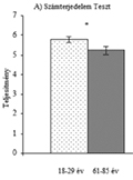
[Changes of cognitive functions in healthy aging]
[Introduction - Mental health has crucial role in our life. Cognitive changes or decline can lead to many difficulties in daily routine of older people (e.g. organization of daily activities), which can, consequently, influence their well-being. Therefore it is an important question, which cognitive abilities are affected by age-related decline. Methods - In our study we aimed to investigate the changes of cognitive abilities in healthy older adults between 61 and 85 years of age compared to the performance of younger adults. Digit span, counting span, listening span, letter fluency, semantic fluency and action fluency tests were used to assess cognitive abilities, namely working memory and executive functions. Results - The results showed that younger adults performed significantly better in all tests than older adults. Importantly, the performance of older adults was better on tests requiring less complex mental computations (e.g. digit span test) than on more complex tests where both storing and mani-pulating information was required (e.g., counting span test). We also showed that within the older age group, cognitive functions’ decline was linearly associated with increasing age. Conclusion - The present study used several, well-established neuropsychological tests to map the changes of working memory and executive functions in healthy older adults between 61 and 85 years of age compared to younger adults. Our findings can contribute to the development of prevention programs aimed at improving the quality of life of older adults and preventing age-related cognitive decline.]
Clinical Neuroscience
MARCH 30, 2018

[Schizotypal traits and verbal creativity]
[Introduction - The relationship of schizophrenia and its milder, subclinical forms, with creativity has been in the centre of theoretical interest for decades, however, the systematic research of the topic only prevailed more recently. Purpose - Here we aimed to examine the connection between different schizotypal and non-schizotypal personality traits and verbal creativity in a nonclinical sample. We also investigated the correspondence of two schizotypy inventories, the Oxford-Liverpool Inventory of Feelings and Experiences and a special character configuration of the Temperament and Character Inventory associated with schizotypy. Method - 57 healthy adults (14/43 m/f, mean age 21.51±1.43 years) - took part in the experiment. All participants received a detailed information sheet and gave informed consent prior to participation. Participants completed the Oxford-Liverpool Inventory and the Temperament and Character Inventory to measure both schizotypal and non-schizotypal personality traits. Torrance Test of Creative Thinking was used to measure verbal creativity. Associations between reported measures were examined with correlational and regression analyses. Results and conclusion - Out of the specific Temperament and Character configuration associated with schizotypy (low self-directedness, cooperativeness and high self- transcendence), we only found low self-directedness to be correlated significantly with Oxford-Liverpool schizotypy rates (Self-directedness-schizotypy: r=-.730, p<.01). There was no significant connection between schizotypal traits and verbal creativity. In our sample, the Self-directedness and Reward-dependence character and temperament subscales predicted significantly the verbal creativity level (Self-directedness: b=.330, p=.015; Reward - dependence: b=-.260; p=.049). Based on our results, besides schizotypal traits, other personality measures might be considered in relation to verbal creativity, providing further details to the empirical investigation of creativity. We found low self-directedness to be correlated with Oxford-Liverpool schizotypy rates, however, the sample size was not large enough to test the concurrent validity of the two inventories. Future studies might consider to extend the study sample, preferably to both clinical and non-clinical populations.]
Clinical Neuroscience
MARCH 30, 2018
[Perinatal stroke - from symptoms to follow-up]
[Background and purpose - We aimed to analyze patient characteristics of term neonates with the diagnosis of stroke between 2006 and 2017 at the 3rd level Neonatal Intensive Care Unit of the Szent János Hospital. Method - We conducted a retrospective and prospective analysis including 18 newborns with stroke. Presentation, imaging methods, etiology and clinical context were discussed. All patients had a follow-up at 2 years of age or later. Subject of the study - In the past 10 years 17 term born and one premature neonate born at 36 weeks of age were diagnosed with stroke in our unit. All patients were born at good condition generally with high Apgar scores (9±1). Cesarean section was performed in 4 cases. Results - With an estimated incidence of one in 1600-4000 births, the incidence of perinatal stroke in our unit was found to be the same as mentioned in the international databeses. Regarding imaging method, cranial ultrasound scan do not visualise arterial ischaemic stroke therefore head MRI is recommended. Neurological symptoms of the patients presented in the first two days of life. Etiology included thrombophilia (4/18), infection (4/18), vascular malformation (2/18), moderate asphyxia (2/18) and pre-eclampsia (2/18). Middle cerebral artery was involved in 50% while the anterior cerebral artery was affected in 33%. The stroke occured in the left hemisphaerium in 44%, in the right side in 39% and was bilateral in 17%. In two cases the stroke was diagnosed in utero. Early childhood developmental support resulted in average or above average gross and fine motor development and cognitive outcome. Conclusion - Presenting neurological symptoms tipically occur in the first few days after birth when perinatal stroke need to be considered among the broad spectrum of neonatal illnesses. Normal developmental outcome can be achieved even in cases of extensive brain damage with early childhood developmental support. Severely impaired development was observed in the cases of in utero stroke. Inherited prothrombotic disorders may have implications for subsequent pregnancies of the mother. ]
Clinical Neuroscience
MARCH 30, 2018
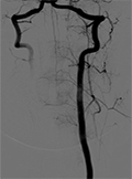
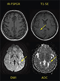
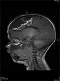
A case with angiographic demonstration of isolated anterior spinal artery occlusion
Anterior spinal artery syndrome (ASAS) is a rare syndrome which occurs due to thrombosis of anterior spinal artery (ASA) which supplies anterior two thirds of the spinal cord. A 27-year-old female patient was admitted to emergency clinic with sudden onset neck pain, sensory loss and weakness in proximal upper extremities which occurred at rest. Thrombophilia assessment tests were negative. Echocardiography was normal. Serum viral markers were negative. In cerebrospinal fluid (CSF) examination, cell count and biochemistry was normal, oligoclonal band was negative, viral markers for herpes simplex virus (HSV) type-1 and type-2, Brucella, Borrellia, Treponema pallidum, Tuberculosis were negative. Diffusion restriction which reveals acute ischemia was detected in Diffusion weighted MRI. Digital subtraction angiography (DSA) was performed. Medical treatment was 300mg/day acetilsalycilic acid. Patient was discharged from neurology clinics to receive rehabilitation against spasticity.
Clinical Neuroscience
MARCH 30, 2018
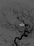
A rare aetiology of stroke; myxomatous aneurysm caused by atrial myxoma
Atrial myxoma is a rare cause of stroke. In this report we present the case of a 52-year-old female patient who went to hospital suffering from a headache. Her neurological examination was normal except for a positive Babinski sign on the left. In the superolateral of the right, a Sylvian fissure consistent with a thrombosed aneurysm was detected using computerised tomography (CT). Diffusion MRI showed an acute infarction on the right MCA area. Transthorasic Echocardiography and ECG were normal. A -16×4 mm-sized fusiform perpendicular aneurysm on the M2 segment Sylvian curve of right MCA and a -6×4 mm-sized dissecting aneurysm on P3 segment of the left posterior cerebral artery (PCA) were observed in cerebral angiography. Transesophageal echocardiography (TEE) demonsrated a large mass with a suspected size of 2×2×1.5 cm on the left atrium. The mass was resected and on the eighth day after the operation, she had a temporary vision loss and hyperintensity on the T1 sequence was interpreted as laminary necrosis suspected on Cranial MRI. In follow up, she was stable with 300mg acetylsalicylic acid treatment. The main treatment is surgical resection in stroke caused by atrial myxoma.
1.
Clinical Neuroscience
Is there any difference in mortality rates of atrial fibrillation detected before or after ischemic stroke?
2023 27. NOV
2.
Clinical Neuroscience
Factors influencing the level of stigma in Parkinson’s disease in western Turkey
2023 27. SEP
3.
Clinical Neuroscience
Neuropathic pain and mood disorders in earthquake survivors with peripheral nerve injuries
2024 03. APR
4.
Journal of Nursing Theory and Practice
[Correlations of Sarcopenia, Frailty, Falls and Social Isolation – A Literature Review in the Light of Swedish Statistics]
2023 31. OCT
5.
Clinical Neuroscience
[Comparison of pain intensity measurements among patients with low-back pain]
2024 03. APR
1.
2.
Clinical Neuroscience Proceedings
[A Magyar Stroke Társaság XVIII. Kongresszusa és a Magyar Neuroszonológiai Társaság XV. Konferenciája. Absztraktfüzet]
2024 06. SEP
3.
4.
Journal of Nursing Theory and Practice
[A selection of the entries submitted to the literary contest "Honorable mission: the joys and challenges of our profession" ]
2024 02. SEP
5.
Journal of Nursing Theory and Practice
[End of Life and Palliative Care of Newborns in the Nursing Context]
2024 02. SEP