The eLitMed.hu medical portal uses computer cookies for convenient operation. Detailed information can be found in the Cookie-policy.
Clinical Neuroscience - 2007;60(05-06)
Content
Clinical Neuroscience
MAY 20, 2007
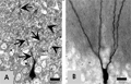
[NOVEL CELL-BIOLOGICAL IDEAS DEDUCIBLE FROM MORPHOLOGICAL OBSERVATIONS ON “DARK” NEURONS REVISITED]
[The origin, nature and fate of ”dark“ (dramatically shrunken and hyperbasophilic) neurons are century-old problems in both human and experimental neuropathology. Until a few years ago, hardly any cell-biological conclusion had been drawn from their histological investigation. On the basis of light and electron microscopic findings in animal experiments performed during the past few years, my research team has put forward novel ideas concerning 1. the nature of ”dark“ neurons (malfunction of an energystoring gel-structure that is ubiquitously present in all intracellular spaces between the ultrastructural elements), 2. the mechanism of their formation (non-programmed initiation of a whole-cell phase-transition in this gel-structure), 3. their capability of recovery (programmed for some physiological purpose), 4. their death mode (neither necrotic nor apoptotic), and 5. their relationship with the apoptotic cell death (the gel structure in question is programmed for the morphological execution of ontogenetic apoptosis). Based on morphological observations, this paper revisits these ideas in order to bring them to the attention of researchers who are in a position to investigate their validity by means of experimental paradigms other than those used here.]


Clinical Neuroscience
MAY 20, 2007

[SLEEP DISORDERS IN PARKINSON SYNDROMES]
[About 90% of neurodegenerative diseases with parkinsonism are associated with sleep disorders including daytime sleepiness, sleep-related breathing disorders and parasomnias. It is hard to define what ratio of insomnia and daytime hypersomnia is caused by the antiparkinsonian tratment, by the somatic and mentalemotional symptoms of the neurodegenerativ disease and by the neurodegenerative brain process itself. Recent research suggests that the latter group is more important than expected. In Parkinson syndromes the structures included in sleep regulation - mainly within the brainstem - are also affected resulting in specific sleep disorders being the primary biological symptoms of these diseases. The recently described parasomnia - REM sleep behavior disorder - has a specific significance in this respect: it may prevent by several years a high ratio of the parkinsonian disorders - especially synucleinopathies - offering the possibility of prevention by identifying the affected individuals. There seems to exist a similar although less clarified association between daytime sleepiness and Parkinson disease. Analysing the behavior of the orexin system in neurodegenerative diseases may help to learn more about this, recently described neurohumoral system and may clear the association of narcolepsy with neurodegeneration. By understanding the associations of parkinsonian disorders and sleep disorders new therapeutical strategies may be invented and may offer new aspects to understand the mechanism of them.]
Clinical Neuroscience
MAY 20, 2007

[DIETARY ASPECTS OF EPILEPSY]
[The ketogenic diet has been used long for the treatment of epilepsy. The high-fat, low-carbohydrate diet creates ketosis. Although the exact mechanism of action is unknown the results are consistent. Ten percent of the patients who start the diet become seizure free, 50% experiences 50% seizure reduction. The diet is hardly tolerable but also effective in therapyresistent patients. Trials are under way with new methods, like the Atkins diet, low-glycaemic-index treatment, polyunsaturated fatty acids, to make the diet more tolerable and widely available.]
Clinical Neuroscience
MAY 20, 2007

[THE PREVALENCE OF WHITE MATTER ABNORMALITIES ON MAGNETIC RESONANCE IMAGES IN MIGRAINE]
[Introduction - While examining patients with headache, abnormalities of unknown significance may quite frequently be encountered. In migraine small, subcortical, white matter abnormalities (WMAs) can be visualized by magnetic resonance images. The connection of these WMAs with the migraine is unclear, but some studies report the higher incidence of WMA in migraine. Patients and method - The authors reviewed the MR scans of their new migraine patients younger than 55 years treated in period of 15 months, and compared the data with a control group. Results - The prevalence of WMA was 10.3% among the migraineurs (78 patients without comorbidities such as hypertension, atherosclerotic heart disease, diabetes mellitus, autoimmun disorder or demyelinating disease) and it was 3.1% in the group of controls (32 persons younger then 55 years, and without migraine or other disease mentioned above). There were patients with WMA both below and above the age of 40; all of them were suffering from migraine without aura with 1 or more attack per month in variable times; none of them had smoked, the majority hadn't used oral contraceptive, and only a few of them used triptan or ergotamin. Conclusion - The data presented here shows that there is a relationship between migraine and WMA. The association of WMA and the risk of following stroke is not cleared. There are well known studies analysing the prevalence of silent infarction too, but we need a long prospective study to answer this question exactly.]
Clinical Neuroscience
MAY 20, 2007
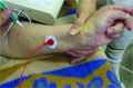
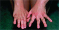
[TREATMENT OF SPASTIC UPPER LIMB WITH BOTULINUS TOXIN]
[Objective - Examination of the effect of local botulinus toxin treatment on spastic upper limb, on patients with different brain injury. Patients and method - Prospective study in Traumatic Brain Injury Rehabilitation Unit of the National Institute for Medical Rehabilitation in the year 2003 and 2004. Thirteen patients (eight with stroke and five with traumatic brain injury) were treated locally on the spastic upper limb with 100 units botulinus A toxin. Results - Spasticity decreased one or two level on Modified Ashworth Scale, and in nine cases the good result were observed still at the end of 3rd month. No local or other complication was detected. Conclusions - Local treatment with botulinus toxin is an effective and safe method to decrease spasticity on upper limb in patients with different brain injury.]
Clinical Neuroscience
MAY 20, 2007

[LATE CONTRALATERAL EPILEPTOGENESIS AFTER INCOMPLETE SURGERY IN TEMPORAL LOBE EPILEPSY FOLLOWED ACROSS 18 YEARS]
[Objectives - To present evidence of changes in seizure semiology suggesting late contralateral epileptogenesis after incomplete surgery in a patient with temporal lobe epilepsy. Methods - The presently 36 year old female patient was followed across 18 years by clinical observation and EEG, and video-EEG monitored before and 18 years after surgery. Results - The patient had complex partial seizures defined by video-EEG which started from the right temporal lobe with an ictal spread to the contralateral (left) temporal lobe. After right amygdalo-hippocampectomy she did not become seizure free. Years after surgery a new type of seizure emerged. Video-EEG monitoring 18 yrs after surgery revealed two seizure types. One started in the right temporal region clinically resembling to the earlier seizures. The new seizure type showed left sided electroclinical pattern. The postoperative MRI detected bilateral hippocampal sclerosis. Side specific memory tasks revealed bilateral hippocampal dysfunctions with subdominant (right) side predominance. Conclusions - The well documented evolution from unilateral to bilateral seizures suggests late contralateral epileptogenesis in which the persisting seizure spread from the primary epileptogenic side and/or the earlier silent contralateral hippocampal sclerosis (HS) may play role. This case show that progressive changes with bilateral involvement may occur during the course of chronic temporal lobe epilepsy.]
Clinical Neuroscience
MAY 20, 2007
[CONGENITAL CATARACTS FACIAL DYSMORPHISM NEUROPATHY SYNDROME - FIRST HUNGARIAN CASE REPORT]
[The congenital cataracts facial dysmorphism neuropathy (CCFDN) syndrome (OMIM 604168) is a recently described autosomal recessive developmental disorder. It is almost completely restricted to an endogamous group of the European Vlax Roma population, called the Rudari. The CCFDN syndrome is a complex phenotype involving multiple systems, characterized by facial dysmorphism, congenital cataracts, microcorneae, delayed early motor and intellectual development, hypogonadotrop hypogonadism, hypomyelination of the peripheral nervous system, and serious complications related to general anaesthesia. This disorder is caused by a homozygous mutation of the carboxy-terminal domain phosphatase 1 (CTDP1) gene, localized to the 18q23 region. Authors present one genetically identified case in a large Roma family. The case documents that the CCFDN mutation is present also in the Hungarian Roma population. Underlie of antropomorphological data the authors presume that the CCFDN mutation reached Hungary as a result of emigration of Vlax Gypsies in the 18th century. The paper calls attention to the fact that molecular genetic diagnostics can replace invasive methods and makes possible the identification of heterozygotes without clinical symptoms. The introduction of the genetic screening enables us to perform genetic counselling and prevention in this high-risk population.]
Clinical Neuroscience
MAY 20, 2007

[PROGRESSIVE MULTIFOCAL LEUKOENCEPHALOPATHY]
[Progressive multifocal leukoencephalopathy is a rare disease caused by the reactivation of an opportunistic agent, JC virus almost in every cases in immunodeficient conditions. The disease is characterized by multifocal demyelinating lesions of the central nervous system and causes death within a few months. The authors report two patients: a 67 year-old male treated because of chronic lymphoid leukemia, and a 19 year-old male having a hereditary immunodeficiency, X-linked hyper IgM syndrome. In both cases continuously progressive right, later both hemispheric signs were detected. Cerebrospinal fluid was not helpful. Brain MRI showed bilateral large, white matter lesion. The progression was not influenced by the treatment, finally both patient died ten and six weeks after the appearance of first complaints. The diagnosis was confirmed by brain biopsy and autopsy in both cases. Our cases demonstrate that progressive multifocal leukoencephalopathy can develop in various immunodeficiencies.]
Clinical Neuroscience
MAY 20, 2007

Clinical Neuroscience
MAY 20, 2007

[100 years of riddle… X. Jubilee Alzheimer’s disease congress on the 100th anniversary of disease description]
[100 years of riddle… X. Jubilee Alzheimer’s disease congress on the 100th anniversary of disease description 2007;60(05-06)]
Clinical Neuroscience
MAY 20, 2007

1.
Clinical Neuroscience
[Headache registry in Szeged: Experiences regarding to migraine patients]
21. MAY
2.
Clinical Neuroscience
[The new target population of stroke awareness campaign: Kindergarten students ]
21. MAY
3.
Clinical Neuroscience
Is there any difference in mortality rates of atrial fibrillation detected before or after ischemic stroke?
27. NOV
4.
Clinical Neuroscience
Factors influencing the level of stigma in Parkinson’s disease in western Turkey
27. SEP
5.
Clinical Neuroscience
[The effects of demographic and clinical factors on the severity of poststroke aphasia]
18. JUL
1.
2.
Clinical Oncology
[Pancreatic cancer: ESMO Clinical Practice Guideline for diagnosis, treatment and follow-up]
29. AUG
3.
Clinical Oncology
[Pharmacovigilance landscape – Lessons from the past and opportunities for future]
29. AUG
4.
5.