The eLitMed.hu medical portal uses computer cookies for convenient operation. Detailed information can be found in the Cookie-policy.
Clinical Neuroscience - 2002;55(07-08)
Content
Clinical Neuroscience
AUGUST 20, 2002

[Parkinson's syndrome and cognitive disorders]
[The cognitive (executive) ability of patients with Parkinson’s-disease (PD) deteriorates gradually during the progression of the disease. Fluency of speech, word finding, working memory, ability to plan the future and flexibility decline. Cognitive disturbance was found to be proportional with the speech, posture, gait and balance problems and can not be influenced by L-dopa substitution. Apart the dorsal and ventral mesolimbic dopaminergic systems the coerulo-cortical noradrenergic, serotoninergic and cholinergic systems are also impaired in PD. Subcortical dementia in PD can also be explained by the functional dysability of dorsolateral and anterior cingular circuits. Attention deficit can be explained by the dopamine depletion of cingular cortex. Cortical Lewy bodies, neurofibrillary tangles, neurit plaques and additional vascular pathology should also play a role in cognitive impairment of PD. In several systemic degenerative diseases associating with Parkinson’s syndrome (PS) ie. progressive supranuclear palsy (PSP), corticobasal degeneration (CBD), multiple system atrophy (MSA) dementia can be detected with various severity, therefore the question arises concerning the correlation between cognitive disability and PS. Parkinson syndrome can also develop in frontotemporal dementias (FTD), Alzheimer’s disease and cortical Lewy body disease (CLBD) but no correlation exists between motor disability and severity of dementia. In CLBD dementia can be the initial symptom in 18% of cases but PS can also preceedes the dementia. In PSP profound depletion of other monoaminergic neurotransmitter system was also reported. In FTDs associated with PS degeneration of substantia nigra, locus coeruleus and basal nucleus of Meynert has been reported with increased number of neurofibrillary tangles. In patients with vascular PS (VP) there is generally no tremor and rigidity, but pseudobulbar palsy, dementia, gate disturbance, incontinency appeares; L-dopa treatment is generally ineffective. In VP no cellular loss can be found within the substantia nigra, but leukoaraiosis, lacunae in the white matter and basal ganglia are commonly demonstrated.]


Clinical Neuroscience
AUGUST 20, 2002

[Investigation of the dopamine dysregulation hypothesis of schizophrenia with neuroimaging techniques]
[The most elaborated biochemical concept of schizophrenia is the dopamine hypothesis. However, this classical theory is based on indirect observations. It has recently become possible to study this theory directly by means of advanced functional neuroimaging techniques, the development of specific radioligands and study protocols that are eligible to monitor dynamic changes in the neurotransmitter systems. According to the early concept, the essence of schizophrenia is the hyperactivity of the dopamine system. Nevertheless, this idea has gone through many modifications. In accordance with the modified dopamine hypothesis, the cognitive deficit and negative symptoms are related to the hypoactivity of the dorsolateral prefrontal cortex while the acute phasis of the disease associates with hyperactivity of the ventral striatal elements of the dopaminergic system. Between these dysfunctions there is causality via their exuberant connections. Beyond that, the interactions between the prefrontal and striatal anomalies implicate the involvement of other neurotransmitters than dopamine. Observations from model psychosis induced by N-methyl-D-aspartate antagonists and in vivo neuroimaging investigations in humans support primarily the role of glutamatergic system. Our developing knowledge about the neurochemical mechanism of schizophrenia can significantly affect therapeutic strategies as well.]
Clinical Neuroscience
AUGUST 20, 2002
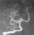
[Cerebellar venous anomalies with symptomatic vascular malformations]
[Cerebral and cerebellar venous anomalies (previously known as venous angiomas) form the alternative venous drainage of the surrounding nervous tissue because of the un-development of the normal venous system. They are made up of veins with abnormal structure: thick walls, lumens dilated and of irregular calibre that converge radially towards a wide draining vein (caput medusae). They are thought to be a benign condition although they are sometimes associated with cerebellar hemorrhages. Authors report three patients with cerebellar venous anomalies associated either with pontine cavernoma, cerebellar arteriovenous malformation or cerebellar infarct. They illustrate that cerebellar venous anomalies are benign conditions, but their presence might be a marker for additional, pathogenic malformation. It might be difficult to detect the associated malformations even by sophisticated imaging methods, but their presence can modify the treatment options.]
Clinical Neuroscience
AUGUST 20, 2002
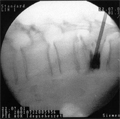
[Percutaneous procedure for treatment of diseased vertebral bodies with different etiology: vertebroplasty]
[Percutaneous vertebroplasty is a radiologically guided invasive technique consisting of the injection of surgical cement into the diseased vertebral body. The procedure results in immediate pain relief and strengthening of the bone due to the polymerization process of the filling material hardening the vertebral body and preventing further collapse. This method is suitable for the treatment of osteoporotic vertebral fractures and of osteolytic vertebral body metastases without neurological signs, in multiple appearance as well. Authors present technical details of the procedure performed by bi-directional fluoroscopy and combined CT-fluoroscopy control as well as short-term experience obtained by treatment of 17 patients.]
Clinical Neuroscience
AUGUST 20, 2002
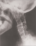
[The molecular genetic control of bony developmental malformations affecting the craniocervical junction and the cervical spine]
[In this review a new interpretation of the origin of bony developmental malformations affecting the craniocervical junction and the cervical spine is presented based on recent advances in the understanding of embryonic development of the spine and its molecular genetic control. Radiographs, CT and MRI scans or CT myelograms of patients with Klippel-Feil syndrome were used for demonstration. Detailed clinical and radiologial analysis of these patients was published earlier [David KM, Stevens JM, Thorogood P, Crockard HA. The dysmorphic cervical spine in Klippel-Feil syndrome: interpretations from developmental biology. Neurosurg Focus 1999;6(6):1.]. Homeotic transformation due to mutations or disturbed expression of Hox genes is a possible mechanism responsible for C1 assimilation. Notochordal defects and/or signalling problems, that result in reduced or impaired Pax-1 gene expression, may underlie vertebral fusions. This, together with asymmetrical distribution of paraxial mesoderm cells and a possible lack of communication across the embryonic mid-line, could cause the asymmetrical fusion patterns. The wide and flattened shape of the fused vertebral bodies, their resemblance to the embryonic cartilaginous vertebrae and the process of progressive bony fusion with age suggest that the fusions occur before or, at the latest, during chondrification of vertebrae. The authors suggest that the aforementioned mechanisms are likely to be, at least in part, responsible for the origin of the bony developmental malformations affecting the craniocervical junction and the cervical spine.]
Clinical Neuroscience
AUGUST 20, 2002

[Neuropsychological outcome following bilateral pallidotomy in patients with Parkinson's disease]
[Introduction - Although significant improvement of motor function following bilateral pallidotomy for the treatment of Parkinsons's disease has been proved, the cognitive sequalae have not been clearly defined. There are recurrent loops interconnecting specific areas of the frontal cortex and the basal ganglia, suggesting the continuity or complementary functioning between these areas. Patients and methods - Pre- and postoperative cognitive function was evaluated in 19 Parkinsonian patients who underwent bilateral pallidotomy in order to clarify its effects on cognitive function. All patients were evaluated one day before the procedure and 12+ months after surgery using neuropsychological tests (Raven Progressive Matrices and Bergen Facial Recognition Test). Proper performance in these tests requires reasoning, abstraction and spatial memory, involving strongly the frontal functions. These functions could be described in terms of the ”working memory” concept. Hand Mental Rotation Test was used as comparing task not involving frontal functions. Scores were analyzed by Student’s t-test. Results - Modest improvement was observed in these cognitive functions as assessed by Raven Progressive Matrices (p<0.0688) and a significant change in the complex parts of Bergen Facial Recognition Test (p<0.0547; p<0.0468) was also noticed, but no change was registered in mental rotation tasks. Conclusion - Present data revealed that bilateral pallidotomy is associated with modest and long-lasting improvement in tasks involving the ”working memory”.]
Clinical Neuroscience
AUGUST 20, 2002
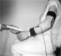
[Application of kinematic parameters for the assessment of impairments due to central motoneuron damage]
[Evidence based medicine requires objective methods for the assessment of status of the patients. The method described by the authors makes it possible to assess motoric impairment of patients in an objective way. It is based on three-dimensional motion analysis. Authors present the case history of two patients with spastic hemiparesis due to central nervous system damage. Changes in motoric impairment were followed by three-dimensional motion analysis. This method can be adapted for the assessment of motor impairment arising from other reasons as well.]
Clinical Neuroscience
AUGUST 20, 2002

Clinical Neuroscience
AUGUST 20, 2002

Clinical Neuroscience
AUGUST 20, 2002
Clinical Neuroscience
AUGUST 20, 2002

1.
Clinical Neuroscience
[Headache registry in Szeged: Experiences regarding to migraine patients]
21. MAY
2.
Clinical Neuroscience
[The new target population of stroke awareness campaign: Kindergarten students ]
21. MAY
3.
Clinical Neuroscience
Is there any difference in mortality rates of atrial fibrillation detected before or after ischemic stroke?
27. NOV
4.
Clinical Neuroscience
Factors influencing the level of stigma in Parkinson’s disease in western Turkey
27. SEP
5.
Clinical Neuroscience
[The effects of demographic and clinical factors on the severity of poststroke aphasia]
18. JUL
1.
2.
3.
4.
5.