The eLitMed.hu medical portal uses computer cookies for convenient operation. Detailed information can be found in the Cookie-policy.
Clinical Neuroscience - 2002;55(05-06)
Content
Clinical Neuroscience
JUNE 20, 2002

[Neurologic complications of aortic dissection]
[Introduction - Beside the damages of the cardiovascular system the lesions of the the nervous system are the most common complications of aortic dissection. This is usually an early event, therefore the dissection of the aorta may manifest itself as an acute primary neurologic disease. The aim of this study is to describe the frequency and distribution of acute neurologic symptoms occurring in aortic dissection and the distribution of their clinico-pathologic features and to establish correlations between these and the acute inhospital mortality as well as to discuss available diagnostic and therapeutic possibilities. Patients and methods - The study was based of 95 cases of acute dissection of aorta (with additional three later events of redissection), observed in a longitudinal study over a period of 29.5 years, in a population of 106 000 (in Western Hungary). Results - Of the 95 patients 20 (21%) died before admission. Neurological complications were observed in 30 of the 75 patients admitted to hospital (40%). Symptoms involving the central nervous system were found in 24 patients, affecting the spinal cord in two and the peripheral nervous system in four cases. The dissection of the aorta was diagnosed in vivo only in 22 out of the 75 patients who died in hospital (29%). 53 patients (71%) without correct diagnosis received supportive therapy only. The average survival time of the 21 patients with proximal dissection of aorta was 48.5 hours. The survival time of 23 patients with the same type of dissection involving the vessels of the aortic arch was 22.2 hours. This difference in survival time was significant (p=0.0152). 20 of 23 patients (87%) in this group showed signs of neurologic damage confirming earlier experience that neurological complications can seriously worsen the otherwise already catastrophic prognosis of aortic dissection. Conclusions - The study brought compelling evidence for the need for early diagnosis and rapid transfer of patients to appropriate cardiac surgery centers for definitive diagnosis and therapy.]


Clinical Neuroscience
JUNE 20, 2002

[Investigation of insertion/deletion polymorphism of the ACE gene on stroke patients]
[Introduction - This is the first Hungarian paper on the insertion/deletion polymorphism of ACE gene in stroke patients. According to literature data, the role of this polymorphism is controversial in the pathogenesis of stroke. The aim was to study the prevalence of the polymorphism in healthy persons and in stroke patients. Patients and methods - Blood samples from 173 unrelated healthy donors and 253 stroke patients were investigated by polymerase chain reaction (PCR). Preivous stroke was documented by CT or MRI and CDS. A routine questionnaire was used to study previous vascular events and the risk profile of patients. Results - I/I allele was found in 20%, I/D 52% and D/D 28% in the healthy group. Prevalence of the pathologic D/D allele did not differ between healthy and patients group (28% and 27%, OR: 0.88, and in subgroup age under 50 years OR: 1.00). No correlation was found between D/D and conventional risk profile but a positiv correlation was found in young patients having D/D and hyperlipidemia (p<0.05) and hyperfibrinogenemia (p<0.05). D/D prevalence was found higher in patients with family anamnesis of myocardial infarction (p<0.05). Very low prevalence of D/D allele was found in cardiogen embolic group (p>0.05). Conclusions - The ACE polymorphism does not seem to be an independent risk factor for stroke. However, in young stroke patients with D/D allele, hyperlipidemia and/or hyperfibrinogenemia present very high risk for stroke.]
Clinical Neuroscience
JUNE 20, 2002

[β-amyloid peptide-induced intracellular calcium level changes in Alzheimer fibroblasts]
[Rationale - β-amyloid peptides, comprising the major neuropathological lesions of Alzheimer's disease, have been found to form depositions in various peripheral tissues, including the skin. Neurons in the disorder succumb to the altered ionic homeostasis and some other factors caused by this toxic peptide. In line with these findings, our study aimed to find differences in biochemical processes of cultured cutaneous fibroblasts derived from sporadic Alzheimer patients and from agematched control individuals that may mirror changes in the central nervous system. Methods - Intracellular ionic homeostasis of Alzheimer and control fibroblasts was measured in Fura-2AMloaded human fibroblasts by dual wavelength spectrofluorimetry. Results - Cells derived from Alzheimer patients exhibited lower intracellular free calcium levels as compared to the control cultures. Exposure of fibroblasts to β-amyloid resulted in increased calcium concentrations of the control cells, but not of Alzheimer ones. Conclusion - Our findings indicate that Alzheimer’s disease is a systemic disorder that, among others, affects the calcium homeostasis of fibroblasts. Even though it is unknown whether the diminished ionic response of Alzheimer fibroblasts is a disease or actual status marker, it could prove to be a useful model for the analysis of Alzheimer specific changes.]
Clinical Neuroscience
JUNE 20, 2002
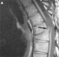
[MR examination of thoracic herniation of the spinal cord]
[Transdural herniation of the spinal cord is thought to be previously extremely rare and very often misdiagnosed. Possible reasons may be iatrogenic and traumatic or in about one third of cases it may be unknown, where the probable origin might be a congenital dural defect. The pathology may show characteristic and misleading MR patterns of the thoracic spine, emphasising the importance of these patterns. This anomaly can lead to progressive Brown-Sequard syndrome. Surgical intervention, consisting the repair of the dural defect may result in improvement or even complete regression of the neurologic deficits.]
Clinical Neuroscience
JUNE 20, 2002
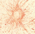
[Identification of gliomas by morphological and immunocytochemical analysis in cell cultures]
[Introduction - The morphology and immunocytochemical properties of 250 different monolayer cultures derived from various human brain tumor specimens were investigated on purpose to support and complement the neuropatholgical diagnosis. In this study analyses of 124 glioma cases are presented. Methods - The tumor samples were mechanically dissociated and seeded on glass coverslips. After the formation of the monolayer cultures were fixed and stained by May-Grünwald- Giemsa method for the morphological examination. Semi-quantitative immunocytochemical labeling included several different types of mono- and polyclonal primary antibodies using avidin-biotin visualization system. In nine cases of the glioblastomas the sufficient proliferation made possible to establish cell lines from the primary cultures. Results - The glial origin of the tumors was identified in 124 cases based upon the presence of glial fibrillary acidic protein. A negative correlation between the intensity of glial fibrillary acidic protein immunostaining and the grade of tumor malignancy was found. During long-term cultivation of the higher grade gliomas the incidence and intensity of glial fibrillary acidic protein labeled cells was decreasing. Both the vimentin and the neuron specific enolase labeling were in general stronger than the glial fibrillary acidic protein and almost all the cells were stained. The incidence of Ki-67 positive cells increased with the grade of malignancy. Concerning the tumor classification our immunocytochemical results correlated with the routine histopathological examination. Conclusions - On the basis of these results we conclude that monolayer cultures obtained from tumor specimens can support and complement the correct diagnosis of the various human brain tumors.]
Clinical Neuroscience
JUNE 20, 2002
[Electrophysiological investigations in Parkinson’s diseae]
[Objective - Post-movement beta synchronization is an increase in EEG beta power after movement termination. Its characteristics in movement disorders are not well described. Tremor dominant Parkinson’s disease shows unique clinical, anatomical and biochemical features. In our study we examined the relation between the laterality of tremor and size of post-movement beta synchronisation in tremor dominant Parkinson’s disease. Methods - In a self-paced movement paradigm we measured movement duration and analyzed EEG power changes at movement-reactive beta frequencies. Results - Movement duration was significantly longer in Parkinson-patients than in controls (0.49±0.170 s, 0.35±0.087 s, p=0.013, Mann-Whitney test). There was no difference between the two hands in the control group (0.36±0.078s, 0.34±0.099 s, p=0.207, Wilcoxon-test), while Parkinson patients performed longer movement with their left hand (0.52±0.195 s, 0.46±0.148 s, p=0.049, Wilcoxon), unrelated to the side of tremor. In controls, post-movement beta synchronisation contralateral to the movement was not significantly different after right and left hand movement (108.1±68.21% and 92.1±23.43%, p=0.78 Wilcoxon). In Parkinson patients post-movement beta synchronisation was significantly smaller contralateral to the tremulous hand movement (36.9±47.79%, 104.7±91.42%, p=0.012, Wilcoxon-test). The post-movement beta synchronisation showed anterior shifting in Parkinson-patients. Conclusions - In tremor dominant Parkinson’s disease the asymmetric decrease of post-move beta synchronisation is related to the laterality of tremor rather than bradykinesia. Analysis of this phenomena might provide further insight to the pathophysiology of Parkinson’s disease.]
Clinical Neuroscience
JUNE 20, 2002
Clinical Neuroscience
JUNE 20, 2002

Clinical Neuroscience
JUNE 20, 2002

Clinical Neuroscience
JUNE 20, 2002

Clinical Neuroscience
JUNE 20, 2002

Clinical Neuroscience
JUNE 20, 2002

Clinical Neuroscience
JUNE 20, 2002

1.
Clinical Neuroscience
[Headache registry in Szeged: Experiences regarding to migraine patients]
21. MAY
2.
Clinical Neuroscience
[The new target population of stroke awareness campaign: Kindergarten students ]
21. MAY
3.
Clinical Neuroscience
Is there any difference in mortality rates of atrial fibrillation detected before or after ischemic stroke?
27. NOV
4.
Clinical Neuroscience
Factors influencing the level of stigma in Parkinson’s disease in western Turkey
27. SEP
5.
Clinical Neuroscience
[The effects of demographic and clinical factors on the severity of poststroke aphasia]
18. JUL
1.
2.
Clinical Oncology
[Pancreatic cancer: ESMO Clinical Practice Guideline for diagnosis, treatment and follow-up]
29. AUG
3.
Clinical Oncology
[Pharmacovigilance landscape – Lessons from the past and opportunities for future]
29. AUG
4.
5.