The eLitMed.hu medical portal uses computer cookies for convenient operation. Detailed information can be found in the Cookie-policy.
Hungarian Radiology - 2006;80(01-02)
Content
Hungarian Radiology
MARCH 20, 2006


Hungarian Radiology
MARCH 20, 2006
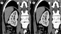
[Diagnostic pitfalls and artifacts of multislice CT]
[There is a spectacular development in diagnostic radiology in the last one and a half decades. State-of-the-art US, CT and MR appliances and the dynamic software developments has improved diagnostic safety by order of magnitude, which resulted in the reduction of possible errors and misinterpretations. The advent of MSCT resulted in shorter scanning times, the submillimeter collimation and the subsecond scan time improves the spatial resolution of the image, the motion artifacts are reduced and the evaluation of the parenchymal organs improves. However, the new technology of MSCT raises new questions. Due to faster data collection the acquisition time decreases, that is why the tracing of the contrast material must be accurately timed. The high contrast material density that appears suddenly in pulsing vessels makes a disturbing effect on its environment, thus making way to erroneous interpretation. The performance of a secondary reconstruction (2D and 3D reconstructions) may diminish the possibility of diagnostic pitfalls and artifacts. Reconstruction increments made from appropriately overlapping thin slices are required for good image quality and spatial resolution, otherwise the image quality is deteriorating, some vessels might “disappear”, they are not depicted. We are struggling with several problems using MIP CTangiography. The proper elimination of the bones, the improper selection of VOI (volume of interest) might lead to false positive result, and the assessment of small vessels might become impossible. The differentiation of soft plaque and vessel thrombus can also be a problem, and the hard plaque may imitate a constriction. The knowledge of breath and pulsating motion artifacts, beam-hardening artifacts and flow-related artifacts is essential. Differentiating difficulties during virtual endoscopy, the partial volumen effect, the interpretation of various post-operative conditions, the disturbing effects of implants may cause diagnostic and differential diagnostic problems. The author gives a summary of possible errors, misinterpretations and artifacts that may occur with the application of MSCT even if examination protocols are followed.]
Hungarian Radiology
MARCH 20, 2006

Hungarian Radiology
MARCH 20, 2006

Hungarian Radiology
MARCH 20, 2006

Hungarian Radiology
MARCH 20, 2006

Hungarian Radiology
MARCH 20, 2006

Hungarian Radiology
MARCH 20, 2006

Hungarian Radiology
MARCH 20, 2006

Hungarian Radiology
MARCH 20, 2006

1.
Clinical Neuroscience
[Headache registry in Szeged: Experiences regarding to migraine patients]
21. MAY
2.
Clinical Neuroscience
[The new target population of stroke awareness campaign: Kindergarten students ]
21. MAY
3.
Clinical Neuroscience
Is there any difference in mortality rates of atrial fibrillation detected before or after ischemic stroke?
27. NOV
4.
Clinical Neuroscience
Factors influencing the level of stigma in Parkinson’s disease in western Turkey
27. SEP
5.
Clinical Neuroscience
[The effects of demographic and clinical factors on the severity of poststroke aphasia]
18. JUL
1.
2.
Clinical Oncology
[Pancreatic cancer: ESMO Clinical Practice Guideline for diagnosis, treatment and follow-up]
29. AUG
3.
Clinical Oncology
[Pharmacovigilance landscape – Lessons from the past and opportunities for future]
29. AUG
4.
5.