The eLitMed.hu medical portal uses computer cookies for convenient operation. Detailed information can be found in the Cookie-policy.
Clinical Neuroscience - 2013;66(01-02)
Content
Clinical Neuroscience
JANUARY 25, 2013
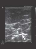
[The significance of high-resolution ultrasonography in the diagnosis of peripheral nerve disorders]
[High resolution ultrasonography is an emerging technique for the investigation of peripheral nerves and is increasingly used worldwide in the diagnosis of peripheral nerve disorders, however, until now it is not widespread in Hungary. According to the literature this method is especially useful in entrapment neuropathies, traumatic peripheral nerve injuries, tumors of the peripheral nerves and sonographically guided interventions. Ultrasonography allows precise morphological analysis and quantitative measurements of the nerves providing useful complementary information to electrodiagnostic data. In entrapment neuropathies ultrasound shows nerve swelling mainly proximal to the sites of compression and a focal change of echotexture. On longitudinal scan, an abrupt caliber change and spindle-like swelling of the compressed nerve segment can be seen. Evaluation of the anatomical background and visualisation of the postoperative and posttraumatic changes provide useful information for planning of the therapy. Ultrasound may be of significant help in localizing the pathological nerve segment when it is at an electrophysiologically inaccessible site or when substantial secondary axonal loss precludes precise electrophysiological localization and it might even show pathological changes when nerve conduction studies are normal. Contrary to electrophysiological investigation ultrasonography might discover neurotmesis in the akute phase of traumatic nerve injuries indicating the necessity of surgical intervention. We provide a summary of the main indications and further application areas of this method.]


Clinical Neuroscience
JANUARY 25, 2013

Clinical Neuroscience
JANUARY 25, 2013

[Aspirin and clopidogrel resistance: possible mechanisms and clinical relevance. Part II: Potential causes and laboratory tests]
[Recent meta-analyses have indicated that patients with vascular disease demonstrated by laboratory tests to be aspirin or clopidogrel-resistant are at an increased risk of major vascular events. The suggested mechanisms of aspirin resistance include genetic polymorphism, alternative pathways of platelet activation, aspirin-insensitive thromboxane biosynthesis, drug interactions, or a low aspirin dose. Clopidogrel resistance is likely to develop as a result of a decreased bioavailability of the active metabolite, due to genetic variation or concomitant drug treatment. Additional work is required to improve and validate laboratory tests of platelet function, so that they may become useful tools for selection of the most appropriate antiplatelet therapy for an individual patient. Improvements in antiplatelet treatment strategies in the future should lead to a reduction in premature vascular events.]
Clinical Neuroscience
JANUARY 25, 2013

[Blood lipid peroxidation, antioxidant enzyme activities and hemorheological changes in autistic children]
[Objectives - Early infantile autism is a severe form of childhood psychiatric disease with characteristic symptoms. Hyperserotoninaemia in 43.5%, lactic acidosis 43% and hyperpyruvataemia in 30% were biochemically demonstrated in autistic children. Our earlier results led to the postulation that a disequilibrium in the blood redox is involved in infantile autism; the oxidative loading and the antioxidant defending enzyme system were investigated together with the hemorheological parameters in infantile autism. Methods - Malonyl-dialdehyde (MDA) endproduct of lipid peroxidation and activities of the antioxidant enzymes: superoxide dismutase (SOD), catalase (C-ase), glutathione peroxidase (GP-ase) and reduced glutathione (GSH) were biochemically determined from plasma and red blood cells. Patients - The antioxidant specificities were investigated in plasma and red blood cell haemolysate from 25 infantile autistic children. Results - Significantly increased superoxide dismutase (SOD) (2.89 vs. 1.32 U/mg protein, p<0.01) and decreased glutathione peroxidase (0.620 vs. 0.910 U/mg protein, p<0.01) levels as well as catalase (0.463 vs. 4.948 BU/mg protein, p<0.001) activities were detected; while the plasma and erythrocyte lipid peroxidation and the reduced glutathione (GSH) levels did not change. The results of the investigated prooxidant and the antioxidant status provide evidence that there exists an oxidative stress in children with infantile autism. While investigating the hemorheological parameters of 25 infantile autistic patients, some characteristic pathological parameters were detected: the initial filtration rate (Fi) (0.72 vs. 0.75 p<0,01) and the clogging rate (CR) (1.926 vs. 2.912, p<0.01) values of red blood cells (RBC) decreased while the mean transit time (Tc) (8.93 vs. 7.39, p<0.001) increased suggesting reduced RBC deformability.]
Clinical Neuroscience
JANUARY 25, 2013
[Experiences with a self developed accelerometer]
[Objective - In neurology the objective evaluation of improvement of paresis on every-day practice. The aim of this study was to develope and test a small 3-d acceleration measuring device and validate its usefulness. Patients and methods - We collected data from 17 mild and medium severity hemiparetic, bedridden acute ischaemic and hemorrhagic stroke patients and compared with data of 22 control subjects. The devices were attached to the paretic and non-paretic extremities and any movements (m/s2) and movement-durations were registered (24h). The data of movement-monitors were compared also with the changes of National Institute of Health Stroke Scale and European Stroke Scale. The electromiograph-sensor of polysomnograph has been used for validation. Results - Mild differences could be found in the use of dominant and non-dominant upper extremities of control persons. The control persons used their upper extremities more frequently than the stroke patients. Our data showed significant correlation with National Institute of Health Stroke Scale. Higher values on the scores were accompanied with less intensive use of extremities. We found a correlation between the consiousness level of patients and their activity of upper extremities. If the patients had severe consiousness disturbances they used significantly less their upper extremities. Conclusion - Our device sensitively detected the movement-differences between paretic and non-paretic extremities and can be used for quantitative evaluation of patient's neurological and consciousness status.]
Clinical Neuroscience
JANUARY 25, 2013

[Intact short-term memory and impaired executive functions in obsessive compulsive disorder]
[Background and purpose - Previous neuropsychological studies produced inconsistent results with tasks tapping short-term verbal and visual-spatial memory and executive functions in obsessive compulsive disorder (OCD). The aim of this study was to investigate the presence of deficits in these cognitive domains. A further goal was to describe the distribution of patients in different impairment ranges for all functions, and clarify the relationship between symptom severity and cognitive impairments. Methods - Thirty patients with OCD (DSM-IV) and 30 healthy volunteers were compared using well-known neuropsychological tasks. We assessed short-term verbal memory with the Digit Span Forward and Digit Span Backward Tasks, short-term visual-spatial memory with the Corsi Block Tapping Task, while we measured the level of executive functions with the StroopTask and the Wisconsin Card Sorting Test (WCST). Results - Compared with a matched healthy control group, the performance of OCD patients was in the impaired range only in the two executive tasks. We find a significant positive correlations between the Y-BOCS (Yale-Brown Obsessive Compulsive Scale) total scores and the number of perseverative responses (r(28)=0.409, p<0.05) and perseverative errors (r(28)=0.385, p<0.05) in the WCST. Conclusion - Our results gave evidence that executive functions are impaired while short-term memory is intact in OCD. This is in line with neuropsychological model of OCD that the deficit of cognitive and behavioral inhibition are responsible for the main cognitive findings of this disorder, most prevalently the deficit in set shifting and prepotent response inhibition.]
Clinical Neuroscience
JANUARY 25, 2013
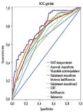
[Early mental test - developing a screening test for mild cognitive impairment]
[Background and purpose - Mild cognitive impairment (MCI) is a heterogenous syndrome considered as a prodromal state of dementia with clinical importance in the early detection of Alzheimer’s Disease. We are currently developing an MCI screening instrument, the Early Mental Test (EMT) suitable to the needs of primary care physicians. The present study describes the validation process of the 6.2 version of the test. Methods - Only subjects (n=132, female 95, male 37) over the age of 55 (mean age 69.2 years (SD=6.59)) scoring at least 20 points on Mini-Mental State Examination (MMSE), mean education 11.17 years (SD=3.86) were included in the study. The psychometric evaluation consisted of Alzheimer’s Disease Assessment Scale Cognitive subscale (ADAS-Cog) and the 6.2 version of EMT. The statistical analyses were carried out using the 17.00 version of SPSS statistical package. Results - The optimalised cut-off point was found to be 3.45 points with corresponding 69% sensitivity, 69% specificity and 69% accuracy measures. The Cronbach-α, that describes the internal consistence of the test was 0.667, which is higher as compared with the same category in the case of the ADASCog (0.446). A weak negative rank correlation was found between the total score of EMT 6.2 and the age of probands (rs=-0.25, p=0.003). Similarly, only a weak correlation was found between the education levels and the total score of EMT 6.2 (rs=0.31, p<0.001). Two of the subtests, the repeated delayed short-time memory and the letter fluency test with a motorical distraction task had significantly better power to separate MCI and control groups than the other subtests of the EMT. Conclusion - The 6.2 version of EMT is a fast and simple detector of MCI with a similar sensitivity-specificity profile to the MMSE, but this version of the test definitely needs further development.]
Clinical Neuroscience
JANUARY 25, 2013
[Sturge-Weber syndrome: clinical and radiological correlates in 86 patients]
[Backgrounds and purpose - To correlate the extent of the leptomeningeal angiomatosis with clinical features in Sturge- Weber syndrome (SWS). Methods - The study group consisted of 86 consecutive patients aged two months to 56 (mean 7.9±10.3) years with SWS and epilepsy. Clinical and MRI data were analyzed. Results - Based on the extent of leptomeningeal angiomatosis, patients were divided into two subgroups: 43 patients had hemispheric angiomatosis and atrophy, whereas, another 43 had focal involvement. Nine of the 43 hemispherial patients (10%) showed bilateral involvement: all of these bilateral cases demonstrated dominance in a single side with hemispheric leptomeningeal angiomatosis and contralateral focal extension. Hemispheric and focal subgroups were clinically different. Patients with hemispheric SWS were younger at the age of epilepsy onset (p<0.001) and age at MRI examination (p<0.05). Neither gender, lateralization, duration of epilepsy, appearance of secondarily generalized seizures, nor seizure frequency revealed a significant difference between subgroups. Conclusion - Bilateral involvement is frequent and occurs in cases with a hemisperic involvement on one side. The age of epilepsy onset is related to the extent of leptomeningeal angiomatosis. Patients with hemispheric form of SWS presented with earlier age of seizure onset. Focal pial angiomatoses do not tend to progress (a longer duration is not associated with more frequent hemispheric involvement). Other variables including seizure frequency and secondary generalized tonic-clonic seizures are not associated with the extent of angiomatosis.]
Clinical Neuroscience
JANUARY 25, 2013

[ASCO classification in clinical practice]
[ASCO (Atherosclerosis, Small vessel disease, Cardiac source, Other cause) is a new of classification of ischemic cerebrovascular diseases. This classification categorizes the data of the patients according to all underlying diseases and allows the clinician to grade the severity of cause (Each of the four phenotypes can be graded 1, 2, or 3). It is suggested to use ASCO classification in large epidemiologic studies but this classification may be used in daily practice. In this study we aimed to analyze the clinical features of patients with ischemic stroke and to investigate results of ASCO classification of these patients and data of 35 patients with ischemic stroke is analyzed. Use of ASCO classification is discussed with the special example cases. Patients' etiology of stroke was classified according to ASCO as known, unknown, completely unknown and unclassifiable group. Percentile of the patients classified as “known” was 71.4% (n=25), “unknown” was 17.1% (n=6), “completely unknown” was 5.7% (n=2) and “unclassifiable group” was 5.7% (n=2). We think that the ASCO classification which is thought to be more useful in large epidemiologic studies may be used in clinical follow-up period of the stroke patients. Further studies, from different neurology centers and stroke units, are needed to expand our experiences about use of ASCO classification in clinical practice.]
Clinical Neuroscience
JANUARY 25, 2013
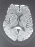
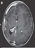
[The presentation of a transient hyperintense lesion with legionnaires disease in a patient, is it a coincidance or an incidental finding?]
[Up to date the presentation of transient splenial lesions in corpus callosum were reported in diffusion weighted magnetic resonance imaging (MRI) only in epileptic patients and patients under antiepileptic therapy. A 41 year old male with no previous medical history was admitted to our clinic with symptoms of pneumonia. The neurological exam revealed stupor, but when awake his speech and orientation were normal. There were no meningeal irritation signs, cranial nerves, piramidal and cerebellar functions were normal. He had moderate respiratory distress and had bilateral rales in lower lobes while oscultating. Laboratory tests revealed high liver function levels and high acute phase reactants. Arterial blood levels showed hypoxemia. A brain MRI showed a hypointensity in the splenium of corpus callosum on T1 weighted images. There was markedly increased signal in this region on diffusion weighted imaging and hypointense on ADC. The lesion was slightly hyperintense on T2 and FLAIR weighted images. A repeat brain MRI was done 30 days after the initial study and showed a complete resolution of the splenial lesion. Transient splenial lesions can be seen due to different mechanisms in different clinical settings. It should be noted that these lesions are mostly reversible. Unnecessary therapies and procedures should be avoided in these lesions.]
Clinical Neuroscience
JANUARY 25, 2013
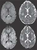
[Mild encephalitis/encephalopathy with a reversible splenial lesion in children]
[Authors, most of them Japanese, have recently published an increasing number of articles on mild encephalitis/encephalopathy with a reversible splenial lesion. We report on two new white European patients and compare published data with our own observations. A 15- year-old girl developed headache, fever, dizziness, vomiting and nuchal rigidity over four days. CSF showed elevated protein and cell count, with the lowest serum Na being 131 mmol/L. MRI on day seven was normal, but she remained febrile, had cerebral edema and episodes of confusion. MRI on day 11 showed a small T2-hyperintense lesion with restricted diffusion in the callosal splenium. Adenoviral infection was proved, and the girl underwent a protracted course of recovery. MRI signal changes improved in six days and disappeared after four months. A 12.5-year-old girl developed headache, lethargy, drowsiness and vomiting. On day five she experienced right-sided numbness, weakness and inability to speak which lasted 12 hours. She was confused and disoriented. MRI disclosed a tiny area of increased T2- signal and restricted diffusion in the splenium. Serum Na was 133 mmol/L, CSF cell count and protein was markedly elevated, and enteroviral infection was detected. Echocardiography showed no changes predisposing to clot formation and no thrombophilia was found. Her symptoms resolved in a week and MRI was normal two months later. These two non-epileptic children increase the small number of white European patients with MERS reported so far. Both had hyponatremia and encephalitis and patient 2 had transient ischemic attack, possibly due to the cerebral edema also resulting in the splenial lesion.]
Clinical Neuroscience
JANUARY 25, 2013

1.
Clinical Neuroscience
[Headache registry in Szeged: Experiences regarding to migraine patients]
21. MAY
2.
Clinical Neuroscience
[The new target population of stroke awareness campaign: Kindergarten students ]
21. MAY
3.
Clinical Neuroscience
Is there any difference in mortality rates of atrial fibrillation detected before or after ischemic stroke?
27. NOV
4.
Clinical Neuroscience
Factors influencing the level of stigma in Parkinson’s disease in western Turkey
27. SEP
5.
Clinical Neuroscience
[The effects of demographic and clinical factors on the severity of poststroke aphasia]
18. JUL
1.
2.
Clinical Oncology
[Pancreatic cancer: ESMO Clinical Practice Guideline for diagnosis, treatment and follow-up]
29. AUG
3.
Clinical Oncology
[Pharmacovigilance landscape – Lessons from the past and opportunities for future]
29. AUG
4.
5.