The eLitMed.hu medical portal uses computer cookies for convenient operation. Detailed information can be found in the Cookie-policy.
Hungarian Radiology - 2004;78(02)
Content
Hungarian Radiology
APRIL 20, 2004


Hungarian Radiology
APRIL 20, 2004
[Staging for lung cancer]
[The author summarizes the current lung cancer staging according to the literature and her more than 10 year experience. This overview is based on the International System for staging lung cancer, which have been adopted by the American Joint Commitee on Cancer and the Union Internationale Contre le Cancer in 1997. TNM classification of lung cancer is based on chest CT and on bronchoscopy in Hungary, since only one PET equipment is available, which is not enough for staging every cancer patient.]
Hungarian Radiology
APRIL 20, 2004
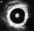
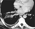
[Endobronchial ultrasonography in the diagnosis of pulmonary and mediastinal malignancies]
[Endobronchial ultrasonography can be performed during conventional bronchofiberoscopy. The main indications are intrathoracic malignancies. Endobronchial ultrasonography has a great signficance in the diagnosis of mediastinal and lung processes and in staging of lung cancer. Endobronchial ultrasound is superior to computed tomography in evaluation of the disorders of tracheal and bronchial wall. The summary about this new modality is based on the international references and the authors’s own experiences.]
Hungarian Radiology
APRIL 20, 2004
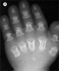
[Opsismodysplasia A report of two cases]
[INTRODUCTION - Opsismodysplasia is a rare, severe, neonatal dwarfism usually associated with fatal outcome in the first few years of life. Up to, 2003 about 15 cases have been reported. CASE REPORT - We describe two brothers six and four years old with opsismodysplasia, who presented to the paediatric orthopaedic clinic with the diagnosis of short stature and genu varum deformity. CONCLUSION - Paediatric specialists should be aware, that in rare instances, with improving medical care, they may see children with severe bone dysplasias which usually do not reach age in which paediatric orthopaedic services are required.]
Hungarian Radiology
APRIL 20, 2004
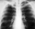
[Necrotising sarcoid granulomatosis: imitator in the chest]
[INTRODUCTION - Both intra and extrapulmonary manifestation of necrotising sarcoid granulomatosis is very rare. It is characterised by variable pulmonary radiological picture, typical histology and benign clinical course CASE REPORT - A young male patient was admitted to our hospital due to a mass lesion in the right apex of the lung suspicious of tuberculosis discovered on plain chest X-ray. Antituberculotic therapy resulted no change of radiological picture. Cytological sample taken by ultrasound guided fine needle aspiration contained no tumor cells. Finally necrotising sarcoid granulomatosis was confirmed by histological examination of the mass removed by atypical surgical resection. CONCLUSION - Intrapulmonary necrotising sarcoid granulomatosis has a variable picture. It has no typical radiological pattern or localisation, therefore it is impossible to diagnose by X-ray morphology. Final diagnosis is based on histology.]
Hungarian Radiology
APRIL 20, 2004

Hungarian Radiology
APRIL 20, 2004

Hungarian Radiology
APRIL 20, 2004

Hungarian Radiology
APRIL 20, 2004

Hungarian Radiology
APRIL 20, 2004

1.
Clinical Neuroscience
[Headache registry in Szeged: Experiences regarding to migraine patients]
21. MAY
2.
Clinical Neuroscience
[The new target population of stroke awareness campaign: Kindergarten students ]
21. MAY
3.
Clinical Neuroscience
Is there any difference in mortality rates of atrial fibrillation detected before or after ischemic stroke?
27. NOV
4.
Clinical Neuroscience
Factors influencing the level of stigma in Parkinson’s disease in western Turkey
27. SEP
5.
Clinical Neuroscience
[The effects of demographic and clinical factors on the severity of poststroke aphasia]
18. JUL
1.
2.
Clinical Oncology
[Pancreatic cancer: ESMO Clinical Practice Guideline for diagnosis, treatment and follow-up]
29. AUG
3.
Clinical Oncology
[Pharmacovigilance landscape – Lessons from the past and opportunities for future]
29. AUG
4.
5.