The eLitMed.hu medical portal uses computer cookies for convenient operation. Detailed information can be found in the Cookie-policy.
Hungarian Radiology - 2004;78(01)
Content
Hungarian Radiology
FEBRUARY 20, 2004


Hungarian Radiology
FEBRUARY 20, 2004
[Theoretical basis of tumor staging The significance of imaging in oncological diseases]
[The general goal for staging patients with any cancer is to determine the overal extent of disease prior to choice of therapy. It has been well documented that biological behavior of any cancer and, therefore, the prognosis of the patient is strongly linked to the extent of the tumor, the presence or absence of lymphatic dissemination as well as systemic metastases. The staging systems have undergone a number of modifications. Classifications of TNM system identifies the depth of tumor invasion (T), the status of regional lymph nodes (N) and the distant metastases. These three parameters are then incorporated into the final clinical stage. There are further modifications which influence the patient survival, e.g. biological, genetical, hystological factors, tumor grade. All cancer cells show dysregulation of cell cycle controll. As the cancer proliferates and the tumor reaches approximately 1-2 mm in diameter further growth recquires the development of new blood vessels (neo-angiogenesis). Intensity of tumor growth has a prognostic influence to the patient's life and depends on the tumor doubling time, which classifies tumors into slow, intermadiate and rapid growing types. The diagnostic impact of imaging is based on the ability of a technique to detect and evaluate the cancer accurately. The very high accuracy and reproducibility of cross-sectional imaging, paticularly computed tomography (CT) and magnetic resonance imaging (MRI) make these methods extremely important in the detection, staging and in the evaluation of the tumors. The revolutionary advances in detection and treatment of malignant disease have led to an increasing role of the radiologist as a member of the multidisciplinary cancer team.]
Hungarian Radiology
FEBRUARY 20, 2004
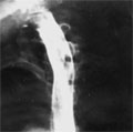
[Candida esophagitis]
[INTRODUCTION - 97-98 percent of upper gastrointestinal mycoses are caused by candida albicans infections. Two cases of candida oesophagitis is reported in female patients with bronchial asthma. CASE REPORT - The patients were treated with steroid drugs due to the asthma. Esophageal barium study in a 48 year old female showed fine multiple rounded radiolucent aphthoid lesions, which fused after six weeks and resulted an aphthoid-shaggy esophagus. In a 55 year old patient double-contrast plain films presented cobblestone, alias snakeskin esophagus and foamy cardia. CONCLUSION - Radiographic signs of candida esophagitis are not pathognomic but correctly demonstrate extension of fungal infection into esophageal wall. Definitive diagnosis can be proved by histological and microbiological examinations.]
Hungarian Radiology
FEBRUARY 20, 2004
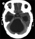
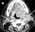
[Recovery of multiple brain abscesses caused by Serratia marcescens in newborn age]
[INTRODUCTION - Multiple brain abscesses caused by Serratia marcescens is a rare disease in newborn infants. The paper describes the development, case history and treatment of the disease. Radiological features of Gram negative bacterial meningitis and brain abscesses are also discussed. PATIENT AND METHODS - A newborn baby boy presented polycythaemia and fever was admitted to the hospital in the first days of his life. On the 11th day after admission convulsions occured and because of suspected intracranial abnormality or meningitis cranial CT was performed. On CT scans multiple abscesses were revealed and surgical therapy including drainage and ventricle shunt was done. During the treatment he had epileptic seisures frequently, but he became symptome free after the introduction of complex antiepileptic therapy. Now the two year old boy is in a good physical condition but he has mild motoromental deficience. CONCLUSION - Radiological imaging plays an important role in the diagnosis and the follow up of brain abscesses and also in the evaluation of its complications.]
Hungarian Radiology
FEBRUARY 20, 2004
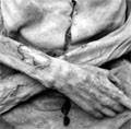
[Pictures to the past Methodological possibilities in the palaeoradiology]
[INTRODUCTION - The radiology as a method is useful not only in the field of the traditional medicine, but in the historic anthropology examining the ancient human remains, and in the palaeopathology examining the pathological changes of the ancient human remains. The aim of our study was the correction of the palaeopathological methods and the radiological diagnosis of the ancient pathological remains. MATERIAL AND METHODS - Approximately 25 specimens originating from the Avar-, the Hungarian Conquest Period and from the Arpadian Age with pathological changes were examined. Besides these 11 naturally mummified individuals were examined by radiological methods, too. The X-ray examinations were done at the Department of Radiology of the National Medical Centre, in Budapest. RESULTS - Vertebral changes, pulmonary tuberculosis and traumatic lesions were found on the mummified individuals. The most frequent changes of the bone alterations were traumas: fractures of the ulna, clavicule, ribs, and trephanation of the skull. Several degenerative changes of the vertebral columns and of the articulation surfaces were detected (osteophyta, spondylosis, arthrosis). The rheumatoid arthritis, Paget-disease, facies leprosa, and the traces of vertebral tuberculosis should be mentioned among the rare pathological disorders. CONCLUSIONS - Since there are no sufficient palaeoradiological methodological references, it was necessary to develop new methods for X-ray examinations.]
Hungarian Radiology
FEBRUARY 20, 2004

Hungarian Radiology
FEBRUARY 20, 2004

Hungarian Radiology
FEBRUARY 20, 2004

Hungarian Radiology
FEBRUARY 20, 2004

Hungarian Radiology
FEBRUARY 20, 2004

Hungarian Radiology
FEBRUARY 20, 2004
1.
Clinical Neuroscience
[Headache registry in Szeged: Experiences regarding to migraine patients]
21. MAY
2.
Clinical Neuroscience
[The new target population of stroke awareness campaign: Kindergarten students ]
21. MAY
3.
Clinical Neuroscience
Is there any difference in mortality rates of atrial fibrillation detected before or after ischemic stroke?
27. NOV
4.
Clinical Neuroscience
Factors influencing the level of stigma in Parkinson’s disease in western Turkey
27. SEP
5.
Clinical Neuroscience
[The effects of demographic and clinical factors on the severity of poststroke aphasia]
18. JUL
1.
2.
3.
4.
5.