The eLitMed.hu medical portal uses computer cookies for convenient operation. Detailed information can be found in the Cookie-policy.
Clinical Neuroscience - 2005;58(11-12)
Content
Clinical Neuroscience
DECEMBER 10, 2005
[THE ROLE OF EVOKED POTENTIAL METHODS IN THE NEUROLOGICAL CLINICAL PRACTICE]
[The author provides an overview on the value of evoked potential (EP) methods (VEP, SEP, BAEP, MEP) in the diagnosis and follow-up of various neurological diseases (multiple sclerosis, cerebrovascular disorders, degenerative diseases, coma, epilepsy, migraine) by reviewing the literature supported by his own clinical experience. While in the past EP was mainly used for establishing the diagnosis, recently, with the expansion of neuroradiology, it has gained a wider use in the assessment of the severity and extent of the pathologic process and especially in longitudinal follow-up. Its role in the diagnostic phase has diminished. In patients with multiple sclerosis the abnormality of the evoked potentials correlate better with the clinical state than with the MRI results. The method is also suitable to monitor the response to therapy. The importance of the EP tests is illustrated by several case demonstrations.]


Clinical Neuroscience
DECEMBER 10, 2005
[EVENT-RELATED EEG AND EVOKED POTENTIAL INVESTIGATIONS IN THE CLINICAL PRA]
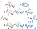
[Considering the limits of the traditional EEG techniques the authors review the main methods and clinical importance of the event-related EEG investigations. According to methods, these can be classified into the spectral analysis of task-related, pre-task and post-task recordings as well as stimuluscontrolled measurements based on evoked potential techniques. The main results of clinical studies on the eventrelated EEG methods are summarized according to chief disease groups (Alzheimer’s disease, epilepsy, schizophrenia, Parkinson's disease, dyslexia, depression). The authors discuss the stimulus-dependent EEG discharges (P300, cognitive potential) in detail. They present the metaanalysis of 224 recent publications on human application of these methods. They analyze the involved scientific areas and the frequency by which these methods were applied in each. Following this, the results of 83 selected clinical studies are summarized. The frequency of the application of the various event-related EEG methods and the tested wave components and other parameters are listed. Finally a summary of the main clinical results is presented again by groups of diseases (schizophrenia, behavioral disorders, traumatic lesions, enuresis nocturna, depression, memory disturbance and dementia, drug effect). Finally, the potential perspectives and the limitations of the event-related EEG methods are briefly discussed.]
Clinical Neuroscience
DECEMBER 10, 2005
[PSYCHOPHYSIOLOGICAL AND CLINICAL ASPECTS OF THE EEG SYNCHRONIZATION RELATED TO COGNITIVE PROCESSES]
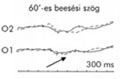
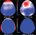
[The authors review the various forms of EEG-synchronization with special emphasis on the characteristics of the induced and enhanced rhythms. The suggested role of the various EEG frequency bands in the cognitive processes is demonstrated by examples from the literature. The relationship between linear and nonlinear electrophysiological complexity and EEG synchronization is analyzed, with a touch on the use of Omega-complexity and synchronization likelihood methods. In the present study the EEG recorded during adding and subtracting tasks was analyzed with the above methods. It was found that during the adding task the theta band increased in the frontal area, which may be related to activation of working memory processes. Mapping the scalp-distribution of synchronization likelihood also confirmed increased synchronization in the frontal area in addition to which increased values were found in the left temporo-parietal area. The analysis of linear and nonlinear EEG synchronization associated with cognitive processing is suitable to explore the task-related and region specific features of these events.]
Clinical Neuroscience
DECEMBER 10, 2005

[ELECTROMYOGRAPHY AT THE BEGINNING OF THE 21st CENTURY]
[This review article is concerned with the role of electromyography (EMG) in the clinical diagnostic work. After a summary on the developmental history of electromyography, the most important EMG methods are presented. The modern quantitative EMG methods are sensitive and accurate thus providing important information in the evaluation of various neurological diseases, particularly in the diagnosis of neuromuscular disorders. The EMG examinations are useful tool for the clinician only if the applied methods are carefully chosen and properly performed and the rules of interpretation are strictly followed.]
Clinical Neuroscience
DECEMBER 10, 2005
[TRANSCRANIAL MAGNETIC STIMULATION: PHYSIOLOGY AND APPLICATIONS]
[Transcranial magnetic stimulation (TMS) is a relatively new technique that allows painless activation of cortical motor neurons. In the clinical setting, TMS is primarily used for the investigation of the corticospinal tract in various neurological diseases, being especially useful in the detection of subclinical dysfunction. In addition to the motor cortex, TMS can be applied to examine other structures inaccessible to electrical stimulation, such as the canalicular portion of the facial nerve. In healthy individuals, TMS can be utilized to monitor excitability changes of the motor system in various situations and muscles, providing valuable information to the understanding of the physiology of motor control. Furthermore, TMS can be used to explore interhemispheric connections as well as intracortical inhibitory and excitatory processes both in health and disease. Finally, with the help of TMS cortical maps of the representation areas of muscles can be constructed, giving insight to both short and long-term cortical plasticity and to the reorganisation of the motor cortex following damage to the brain or acquisition of new motor skills]
Clinical Neuroscience
DECEMBER 10, 2005
[BASIC NEUROGRAPHY AND ITS DIAGNOSTIC IMPORTANCE]
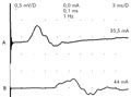
[Nerve conduction studies are fundamental elements of the neurophysiological investigation of neuromuscular diseases. They provide information on peripheral nerve function. Knowledge of the biological and technical basis of the method is essential for the clinician to understand the place of nerve conduction testing in the diagnostic process. A characteristic feature of the nerve fibers is their ability to conduct electrical potentials. This conductivity changes in pathologic circumstances; therefore, the patient's nerve conduction data may be important if a neuromuscular disorder is suspected. The electrical activity spreading along the nerve fibres can be detected with special techniques and instruments. To perform an examination, a stimulator, a high quality amplifier and a computer with various accessories are necessary. The examination is usually carried out by surface stimulation and recording electrodes and requires some cooperation. By supramaximal stimuli all nerve fibers in the peripheral nerve are activated, and their summated activity is recorded bipolarly. For technical reasons the procedures for the motor and the sensory nerve conduction measurements are somewhat different, but their principles are similar. A number of parameters, such as the latency, the amplitude, the area and the shape of the evoked potentials are analyzed. These parameters are influenced by many biological (age, gender, body height, etc.), physical (such as limb temperature) and technical factors. The results are compared with the reference data. Nerve conduction studies may help distinguish between normal and diseased nerve function. The latter has two main categories; axonal lesion and demyelinisation. Axonal lesion is characterized by relatively normal conduction velocity and lower than normal amplitude of the potentials. Demyelinisation is almost the opposite with long latencies, slow conduction velocity and relatively spared potential amplitudes. Nerve conduction studies help differentiate between these two forms. Abnormalities found by nerve conducion measurement may reflect the severity of the disease. Repeated studies are suitable for quantitative follow-up. The anatomical, physiological, pathophysiological and technical details are discussed below. The characteristic neurographic findings of various diseases are also summarized.]
Clinical Neuroscience
DECEMBER 10, 2005

Clinical Neuroscience
DECEMBER 10, 2005

1.
Clinical Neuroscience
[Headache registry in Szeged: Experiences regarding to migraine patients]
21. MAY
2.
Clinical Neuroscience
[The new target population of stroke awareness campaign: Kindergarten students ]
21. MAY
3.
Clinical Neuroscience
Is there any difference in mortality rates of atrial fibrillation detected before or after ischemic stroke?
27. NOV
4.
Clinical Neuroscience
Factors influencing the level of stigma in Parkinson’s disease in western Turkey
27. SEP
5.
Clinical Neuroscience
[The effects of demographic and clinical factors on the severity of poststroke aphasia]
18. JUL
1.
2.
Clinical Oncology
[Pancreatic cancer: ESMO Clinical Practice Guideline for diagnosis, treatment and follow-up]
29. AUG
3.
Clinical Oncology
[Pharmacovigilance landscape – Lessons from the past and opportunities for future]
29. AUG
4.
5.