The eLitMed.hu medical portal uses computer cookies for convenient operation. Detailed information can be found in the Cookie-policy.
Hungarian Radiology - 2010;84(03)
Content
Hungarian Radiology
OCTOBER 15, 2010


Hungarian Radiology
OCTOBER 15, 2010

[Emergency imaging in pregnant women]
[The use of adequate imaging protocol reduces morbidity and mortality in emergency cases of pregnant women. The least harmful, but sufficiently informative imaging method should be chosen that provides us with the diagnosis in the shortest delay. Although the reduction of harmful effects is important, life threatening complications can be avoided by an indicated and well effectuated imaging method using ionizing radiation.]
Hungarian Radiology
OCTOBER 15, 2010

[Pulmonary abnormalities in haematological malignancies - The role of imaging in differential]
[Patients with hematological malignancies may develop a wide range of pulmonary abnormalities due to the hematological disease itself as well as in response to therapy. Immunosuppression and intensive chemotherapy induced severe neutropenia hold a high risk of infection. Infectionrelated morbidity and mortality are still high. One of the most common infectious complications is invasive mycosis, which is lethal in a high percentage of cases if not treated immediately and adequately. Non-infectious complications, such as secondary pulmonary lymphoma, thromboembolism, hemorrhage or drug induced fibrosis may develop during the course of the disease. Sometimes it is difficult to make a definitive diagnosis. As invasive methods (bronchoscopy, bronchoalveolar lavage, biopsy) are mostly contraindicated in these patients with severe neutropenia and thrombocytopenia, imaging techniques are especially important. It is a great challenge to differentiate infectious and noninfectious processes. CT and HRCT play an essential role in differential diagnosis. An early and accurate diagnosis is sometimes the only chance for survival.]
Hungarian Radiology
OCTOBER 15, 2010
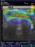
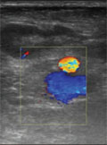
[Differential diagnosis of malignant cervical lymph nodes with real-time ultrasonographic elastography and Doppler ultrasonography]
[PURPOSE - Real-time ultrasonographic elastography is a new imaging technique which is used in characterizing the difference in hardness between pathologic and normal tissue. The purpose of our study was to evaluate the diagnostic performance of real-time ultrasonographic elastography and Doppler ultrasonography (DUS) individually and combined in differentiation of benign and malignant cervical lymph nodes (LN). PATIENTS AND METHODS - Fifty-one patients (12 men, 39 women) referred for fine-needle aspiration or surgical biopsies of suspected cervical lymph nodes were examined with gray scale ultrasonography, power DUS, and realtime ultrasonographic elastography. During DUS examination vascularity and resistance index (RI) values were evaluated. A five-group elastographic colour code pattern was used to evaluate the ultrasonographic elastograms for LN (pattern 1, an absent or a very small hard area; pattern 2, hard area <45%; pattern 3, hard area ≥45%; pattern 4, peripheral hard and central soft areas; pattern 5, hard area occupying entire solid component with or without soft rim). In addition, strains of LN and surrounding muscles were measured on elastograms, and the muscle-to-LN ratio (strain index) was calculated. Real-time ultrasonographic elastography and DUS results were compared with the final diagnosis obtained by fine-needle aspiration cytology analysis and/or by surgical pathology. The diagnostic potential of the examined criteria for malignancy was evaluated with univariate analysis and multivariate generalized estimating equation regression p≤0.05 indicated statistical significance. RESULTS - A strain index higher than 2.45 and colour pattern 4-5 had high utility in malignant LN classification with 93.8% sensitivity, 89.5% specificity (p<0.001). The results were significantly better than those obtained by using DUS characterization - that is, RI greater than 0.57 - which had 78.9% sensitivity (p<0.001). CONCLUSION - Real-time ultrasonographic elastography had 93.8% sensitivity and 89.5% specificity in the differentiation of benign and malignant cervical LN in patients referred for fine-needle aspiration or surgical biopsies with suspicion of malignancy. Real-time ultrasonographic elastography and DUS in addition to gray scale ultrasonography may improve the differential diagnosis of LN.]
Hungarian Radiology
OCTOBER 15, 2010
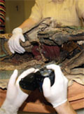
[Interdisciplinary mummy research - Paleoradiology and paleopathology research of the Archbishop of Kalocsa Pál Széchényi’s mummy]
[Purpose - The scientific study of Pál Széchényi’s mummy was carried out by our research team composed of the members of several institutions in 2007 April. The scientific examinations represented a milestone, since up till now it was unclear whether it was a natural or artificial mummy and the century-old question whether Pál Széchényi was in fact a victim of arsenic poisoning in 1710 or it was only a legend could also be answered. Methods - The non-invasive examinations were carried out with multislice CT, traditional X-ray, biopsy, toxicology, energy-dispersive X-ray, X-ray fluorescens analysis, 3D optical digitalization indicator system, endoscope and 3D rapid-prototyping printing. Results - The examinations proved that Archbishop Pál Széchényi’s death was natural, and his body was artificially mummified. The CT examinations were carried out in a slice thickness of under 1 mm facilitating printed skull copy made by 3D rapid-prototyping method which provided the exact and detailed basis for the facial reconstruction.]
Hungarian Radiology
OCTOBER 15, 2010
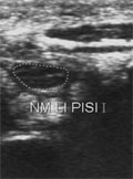
[Fournier’s gangrene]
[INTRODUCTION - Fournier’s gangrene is a rare, rapidly progressive specific form of necrotizing fasciitis that involves the soft tissues of the genital and perianal regions. The infection may spread from these regions along the abdominal wall or towards the lower limbs and has a very high mortality rate. History of the patient, the clinical features and the imaging techniques all together help to recognize the condition and to localize the affected area. CASE REPORT - We present the case of a thirty-year-old male patient, who had on admission a swollen left lower limb. Deep venous thrombosis was excluded by Doppler us. A few days later an other us examination revealed the fasciitis of the left thigh and the scrotum, originating from the deep gluteal bed-sore of the patient. A CT was performed to define the extent of the process and help the planning of the surgical intervention. The patient has recovered well after the operation. CONCLUSION - Due to the high mortality rate of Fournier’s gangrene it is essential to recognize this condition in time and to provide the appropriate surgical intervention or antibiotic therapy. Imaging modalities play a very important role in the diagnosis of Fournier’s gangrene.]
Hungarian Radiology
OCTOBER 15, 2010
[Acute carpal tunnel syndrome after hyperventilation tetany]
[INTRODUCTION - Nerves show the same morphologic changes under compression. CASE REPORT - We describe the morphologic changes of the nerve in temporal course in an acute carpal tunnel syndrome after hyperventilation tetany. CONCLUSION - High-resolution sonography is suited for the imaging of peripheral nerve lesions.]
Hungarian Radiology
OCTOBER 15, 2010

Hungarian Radiology
OCTOBER 15, 2010

Hungarian Radiology
OCTOBER 15, 2010

Hungarian Radiology
OCTOBER 15, 2010

Hungarian Radiology
OCTOBER 15, 2010

Hungarian Radiology
OCTOBER 15, 2010

Hungarian Radiology
OCTOBER 15, 2010

Hungarian Radiology
OCTOBER 15, 2010

1.
Clinical Neuroscience
[Headache registry in Szeged: Experiences regarding to migraine patients]
21. MAY
2.
Clinical Neuroscience
[The new target population of stroke awareness campaign: Kindergarten students ]
21. MAY
3.
Clinical Neuroscience
Is there any difference in mortality rates of atrial fibrillation detected before or after ischemic stroke?
27. NOV
4.
Clinical Neuroscience
Factors influencing the level of stigma in Parkinson’s disease in western Turkey
27. SEP
5.
Clinical Neuroscience
[The effects of demographic and clinical factors on the severity of poststroke aphasia]
18. JUL
1.
2.
Clinical Oncology
[Pancreatic cancer: ESMO Clinical Practice Guideline for diagnosis, treatment and follow-up]
29. AUG
3.
Clinical Oncology
[Pharmacovigilance landscape – Lessons from the past and opportunities for future]
29. AUG
4.
5.