The eLitMed.hu medical portal uses computer cookies for convenient operation. Detailed information can be found in the Cookie-policy.
Hungarian Radiology - 2009;83(03)
Content
Hungarian Radiology
OCTOBER 20, 2009


Hungarian Radiology
OCTOBER 20, 2009
[Role of imaging in the managment of colorectal cancer]
[Colorectal cancer is the third most common cancer worldwide and the second most common cause of cancer death in Hungary. Diagnosis requires the examination of the entire large bowel by means of radiological and/or endoscopic techniques. Colorectal cancer primarily develops from adenomatous polyp over a period of 10-15 years. Tumour staging is crucial for the prognosis and for the planning of the most suitable anticancer therapy. The role of imaging in colorectal cancer is increasing with the change in complex tumour therapy. With advances in ultrasonography (US), computed tomography (CT), and magnetic resonance imaging (MRI) techniques accuracy of imaging has improved. The accuracy of CT improved with the advent of the multislice technique (MDCT). Sensitivity and specificity of CT colonography (CTC) in colon polyps and cancer is over 90%, therefore it is one of the screening tools. Accuracy of the CTC is comparable to the optical colonoscopy, complements conventional colonoscopy well and it is an effective tool in the right hands. Endorectal US (ERUS) depicts the anatomic layers of the rectal wall with high degree of accuracy, therefore it is the best method for the evaluation of the lower tumour stage. High resolution MRI is the most suitable technique for predicting rectal tumor stage, therefore it has been established as the standard for preoperative assessment of rectal cancer.]
Hungarian Radiology
OCTOBER 20, 2009

[Colorectal cancer screening: Lessons from the American experience]
[Colorectal cancer is one of the most common malignancies of our times. The disease takes many years to develop and is typically preceded by polyp formation, which allows timely screening and diagnosis. A number of tests and procedures have been developed to screen for colorectal cancer and its premalignant conditions. However, apparent heterogeneity of the disease, redundancy of the available screening modalities, as well as costs and potential pitfalls of these preventive measures require careful strategic planning. In the United States, recent advances in the campaign for colorectal cancer screening provide important lessons that may guide similar initiatives in Hungary.]
Hungarian Radiology
OCTOBER 20, 2009

[Ways of imaging the expression of vascular endothelial growth factor and its receptor]
[The vascular endothelial growth factor (VEGF) and its receptors (VEGFR) signal-transduction pathway play a key role in the regulation of angiogenesis. It was originally isolated as a selective mitogen for endothelial cells and as a powerful vascular permeability increasing factor. The vascular imaging techniques make the quantification and localization of blood vessels possible. They have been used to assess blood flow, oxygenation, and vascular permeability. Also, they can be used to examine the molecular and cellular difference in the vascular wall. To evaluate tumour vascularity, a multimodality approach is expanding. VEGF as the primary mediator for vascular-permeability is indirectly measurable with DCE-MRI (dynamic contrastenhanced MRI). MRI investigation can determine the ratio of deoxyhemoglobin/oxyhemoglobin in order to localize the hypoxic regions in vivo (BOLD [blood oxygen-level dependent] sequence and OMRI [Overhauser MRI]). In molecular MRI (mMRI), contrast agent-mediated alteration of tissue relaxation times can allow for the detection and localization of molecular disease markers. To localize the expression of VEGFR with SPECT and PET, antibodies and VEGF isoforms can be marked with isotopes. VEGFR is an excellent candidate for targeted ultrasound imaging since it is almost exclusively expressed on activated endothelial cells. Optical imaging is a relatively cheap method suitable so far primarily for small animal studies.]
Hungarian Radiology
OCTOBER 20, 2009
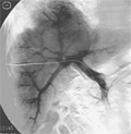
[Portal embolisation prior to liver resection]
[INTRODUCTION - By partial embolisation of the vena portae the number of the patients suitable for radical liverresection can be enhanced, the safety of the operation can be increased, the subsequent results improved. The method is based on the experience that when blocking the circulation of the portal system in special segments of the liver, the other part of the organ tries to substitute the functional deficiency by hypertrophy. Vena portae embolisation is justified in cases when the liver substance remaining after the planned operation is critically small. PATIENTS AND METHODS - The authors carried out vena portae embolisation at Debrecen University Medical and Health Science Centre since October 2003 on six patients. Assessments were made studying the volume of the whole liver, the lobe affected by embolisation and that of the unaffected lobe, by CT-volumetry. The average age of the patients (four men and two women) was 63 years (51-67 years). The hepatic tumour was an extended metastasis localised to one lobe in five cases, and HCC in one of the patients. In each case we carried out closing the right lobe’s portal system. RESULTS - In five cases the left lobe showed increase following the portal embolisation of the right lobe intended to be removed. On the average four-six weeks passed between the two CT-examinations. The growth of the left lobe was an average of 42% (min. 11.8%, max. 75.6%). CONCLUSION - In selected patients the embolisation of the vena portae system of the tumorous liver-segments is a suitable method for enlargening the size of the liver substance remaining after an extensive resection.]
Hungarian Radiology
OCTOBER 20, 2009
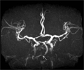
[Interventional neuroradiological treatment of a symptomatic basilar artery stenosis in the background of recurrent brainstem transient ischemic attack]
[INTRODUCTION - Symptomatic stenosis of the basilar artery carries high risk of brainstem stroke. In case of acute thrombotic occlusion of the basilar artery the mortality is over 70%. Besides best medical treatment, angioplasty was employed for 30 years previously, but the results were only acceptable after the advent of flexible intracranial stents. We report our case to call attention to the availibility of this interventional neuroradiological procedure. CASE REPORT - A 64 years old male patient was admitted to our hospital with intermittant diplopia. 90% stenosis of proximal basilar artery was reported by DSA. After balloon dilatation, intracranial stent was placed into the lesion. Symptoms have not recurred three months following the intervention. Control TCD and MR-angiography were performed, and good blood flow ratio was detected in the vertebrobasilar system. CONCLUSION - In the background of recurrent brainstem TIA a basilar artery stenosis has to be searched because the development of a definitive brainstem stroke can be prevented by interventional neuroradiological procedure.]
Hungarian Radiology
OCTOBER 20, 2009
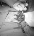
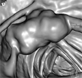
[Omental infarction diagnosed with ultrasonography]
[INTRODUCTION - Omental infarction is a rare entity, mimicking symptoms of acute appendicitis. Although omental infarction has typical morphology, both on sonography and CT, it is rarely diagnosed preoperatively. CASE REPORT - A 4-years-old girl presenting with right lower quadrant abdominal pain underwent abdominal sonography, which revealed normal appendix and a superficial hyperechoic solid mass at the site of the pain. The patient underwent laparotomy which confirmed the presumed diagnosis of partial omental necrosis. The necrotic tissue was resected, the appendix was normal. After uneventful postoperative course the child became symptom -free and was discharged. CONCLUSION - The sonomorphology of omental infarction is typical. The suspicion of this entity should be considered as a differential diagnosis of appendicitis, especially if normal appendix can be visualized.]
Hungarian Radiology
OCTOBER 20, 2009

[Experimental e-Learning course in the further training of Hungarian radiographers]
[The growth in digital media technology within the past decade has strengthened e-Learning’s position in education. Traditional teaching methods are changing: from teacher to moderator and passive learner to active learner. A pilot e-Learning course in MR angiography (MRA) has been undertaken with Hungarian radiographer participants. The e-Learning course consisted of three phases covering basic MRA physics and technical aspects, advanced MRA techniques and concluding with an assessment work. The promising results suggest that e-learning may be a feasable method for the continuing professional development of Hungarian radiographers.]
Hungarian Radiology
OCTOBER 20, 2009

Hungarian Radiology
OCTOBER 20, 2009

Hungarian Radiology
OCTOBER 20, 2009

Hungarian Radiology
OCTOBER 20, 2009

Hungarian Radiology
OCTOBER 20, 2009
Hungarian Radiology
OCTOBER 20, 2009

Hungarian Radiology
OCTOBER 20, 2009

Hungarian Radiology
OCTOBER 20, 2009

Hungarian Radiology
OCTOBER 20, 2009

1.
Clinical Neuroscience
[Headache registry in Szeged: Experiences regarding to migraine patients]
21. MAY
2.
Clinical Neuroscience
[The new target population of stroke awareness campaign: Kindergarten students ]
21. MAY
3.
Clinical Neuroscience
Is there any difference in mortality rates of atrial fibrillation detected before or after ischemic stroke?
27. NOV
4.
Clinical Neuroscience
Factors influencing the level of stigma in Parkinson’s disease in western Turkey
27. SEP
5.
Clinical Neuroscience
[The effects of demographic and clinical factors on the severity of poststroke aphasia]
18. JUL
1.
2.
Clinical Oncology
[Pancreatic cancer: ESMO Clinical Practice Guideline for diagnosis, treatment and follow-up]
29. AUG
3.
Clinical Oncology
[Pharmacovigilance landscape – Lessons from the past and opportunities for future]
29. AUG
4.
5.