The eLitMed.hu medical portal uses computer cookies for convenient operation. Detailed information can be found in the Cookie-policy.
Hungarian Radiology - 2008;82(03-04)
Content
Hungarian Radiology
JUNE 22, 2008


Hungarian Radiology
JUNE 22, 2008
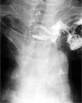
[Complications after subtotal extirpation of the esophagus and the radiological aspects of their treatment]
[INTRODUCTION - Complications after subtotal esophageal extirpation (for eg.: insufficiency of anastomosis, stricture, etc.) and their treatment have special radiological considerations. Surgical departments and their radiologists meet early complications. But, all other radiological departments can face late complications, so the knowledge of these is important for all radiologists. PATIENT AND METHODS - 58 subtotal esophageal extirpations were performed in the last 5 years in the 1st Department of Surgery, Semmelweis University, Budapest. All postoperative radiological examinations were undertaken by the Department of Diagnostic Radiology and Oncotherapy. Most plain chest X-rays and swallowing examinations were made per-os with non ionic water soluble, low osmolarity contrast media on the 7th postoperative day. Further examinations were performed only if needed clinically. RESULTS - The complications were categorized as early and late. Early complications were aspiration, anastomosis insufficiency (31% of cases). Nine cases had self-dilating stents placed because of suture insufficiency. Further complications were noted following stenting (eg.: occlusion, dislocation of stent, closure of stent because of granulation tissues). Amongst late complications, stomach dilatation caused by pylorus-spasm (1 case), stricture of anastomosis (14 cases) and ’too-long’ esophageal stump (3 cases) were seen. CONCLUSION - Conventional radiology has a major role in diagnostics and treatment of complications of esophageal extirpation. Dynamic radiological examination and team work are required between the radiologist and surgeon. Also, it is crucial to cultivate good cooperation in cases of stent implantation, as contrary to the published literature, we find it better to have stents placed under flouroscopy rather than endoscopically.]
Hungarian Radiology
JUNE 22, 2008
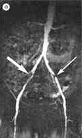
[Significance of MR-angiographic technical parameters and the contrast material in the diagnosis of peripherial vascular diseases]
[Atherosclerosis presents as a significant problem in everyday healthcare. Thus far, its effect on the vascular bed was measured by means of digital subtraction angiography. More recently, due to advance in hardware, the less invasive techniques like the ultrasound, CT and MRI have into the practice, producing results very similar to those of digital subtraction angiography. Recently, contrast material aimed for blood-pool MR angiography has been introduced. This change presents as a challenge to the operating personnel since the injection rate and the MR sequence parameters need to be adjusted adequately. Also, there is a late-phase breakdown of the contrast material. A further challenge is to interpret the steadystate images. In this article an effort has been made to summarise the basis of MR angiography, with special emphasis on peripheral angiography and suitable contrast materials. Finally, we illustrate our parameters through concrete cases.]
Hungarian Radiology
JUNE 22, 2008

Hungarian Radiology
JUNE 22, 2008
Hungarian Radiology
JUNE 22, 2008

Hungarian Radiology
JUNE 22, 2008
Hungarian Radiology
JUNE 22, 2008

Hungarian Radiology
JUNE 22, 2008
Hungarian Radiology
JUNE 22, 2008

Hungarian Radiology
JUNE 22, 2008

1.
Clinical Neuroscience
[Headache registry in Szeged: Experiences regarding to migraine patients]
21. MAY
2.
Clinical Neuroscience
[The new target population of stroke awareness campaign: Kindergarten students ]
21. MAY
3.
Clinical Neuroscience
Is there any difference in mortality rates of atrial fibrillation detected before or after ischemic stroke?
27. NOV
4.
Clinical Neuroscience
Factors influencing the level of stigma in Parkinson’s disease in western Turkey
27. SEP
5.
Clinical Neuroscience
[The effects of demographic and clinical factors on the severity of poststroke aphasia]
18. JUL
1.
2.
Clinical Oncology
[Pancreatic cancer: ESMO Clinical Practice Guideline for diagnosis, treatment and follow-up]
29. AUG
3.
Clinical Oncology
[Pharmacovigilance landscape – Lessons from the past and opportunities for future]
29. AUG
4.
5.