The eLitMed.hu medical portal uses computer cookies for convenient operation. Detailed information can be found in the Cookie-policy.
Hungarian Radiology - 2008;82(01-02)
Content
Hungarian Radiology
MARCH 22, 2008
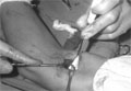
[Osteoid osteoma: diagnosis and treatment]
[Osteoid osteoma is a rare, generally small benign tumor that requires treatment due to the intense pain it causes. The aim of this review is to discuss the basics (pathophysiology, classification and imaging modalities of osteoid osteoma). Further, we have made an attempt to discuss in detail the technique and effects of surgery and radiofrequency ablation in the treatment of osteoid osteoma.]


Hungarian Radiology
MARCH 22, 2008
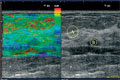
[Initial experiencies with sonoelastography in the examinations of breast diseases]
[INTRODUCTION - Recently the ultrasound examination of the strain of circumscribed breast diseases has been introduced in the non-invasive breast examination, called sonoelastography. In Hungary, the authors had the first possibility to start with this method. They report on their initial experiences. MATERIAL AND METHODS - 61 circumscribed breast lesions in 41 patients sonoelastographic examinations were performed by Hitachi EUB 6500 system using a EZUTE3 real-time elastography unit. 48 lesions strain-ratio was calculated. 22 masses were verified pathologically (18 benign and 4 malignant) and 39 were considered benign upon the findings of clinical mammography and by follow up. They classified them on the basis of patterns published by Itoh, et al. The examinations were done by three experienced radiologists. The classification was done by consensus. RESULT - The all lesions which were verified pathologically or on the basis of examinations or follow ups were thought benign showed the pattern type from 1 to 3. Most of the cystic lesions showed the streaky cystic pattern. There were small number of malignant lesions in their material, and all of them gave the elastic pattern of 4 and 5. The numbers of strain-ratio of zone lesions with pattern 3 overlapping with the lesions of malignant ones. CONCLUSION - The first results showed that both the coloured elastographic pattern and the quantitative strainratio could be used well in the non-invasive diagnostic procedure of breast lesions. It could increase the diagnostic safety. Larger number of examinations are necessitated to find the exact diagnostic role of this method.]
Hungarian Radiology
MARCH 22, 2008
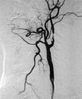
[Use of covered stents in the endovascular treatment of extracranial stenosis of the internal carotid artery]
[INTRODUCTION - Significant stenosis of the internal carotid artery is frequently treated with stent placement. With growing clinical experience and usage of finer instrumentation, the incidence of periprocedural complications have reduced in larger centers. Two-thirds of the complications are postprocedural, due to the embolisation through the stent structure. Covered stents seem to be a good option against such embolisation. Our study demonstrates the efficiency, safety and feasibility of covered stent grafts, and the long term outcome of patients who underwent endovascular treatment of extracranial internal carotid artery stenoses, caused by highly embologenic plaques. MATERIALS AND METHODS - Between 2002 and 2003, 30 patients (22 male, 8 female, aged 50-89yrs, mean: 66 yrs) with 30 internal carotid artery stenoses having ipsilateral symptoms and/or stenotic lesions caused by irregular or ulcerated soft plaques or restenosis were treated with self-expanding covered stents (Symbiot, Boston Scientific). Predilatation and protecting devices were not used. Postdilatation was applied in every patient. Mean followup was 60 months (range 57-66 months), by Doppler ultrasonography as well as clinical examination. RESULTS - The degree of stenosis was found to range from 70% to subtotal occlusion. The plaque surfaces were irregular or ulcerated in 70%. The stenotic lesions were up to 30 mm in length. The narrowing of the internal carotid artery never extended to the common carotid artery. The technical success rate of stenting was 100%. The stents could be positioned with an accuracy of 2-3 mm. Periprocedurally, there were no neurological complications or deaths. During follow-up no strokes or stroke-related deaths occurred. Restenosis was found in two patients (6,6%) who underwent successful balloon dilatation. CONCLUSION - Our experience indicates that the covered stent is an efficient periprocedural and postprocedural “protecting device” to prevent neurological complications due to embolizations caused by high-risk plaques in stenotic lesions of extracranial internal carotid artery.]
Hungarian Radiology
MARCH 22, 2008

[Analysis of the status of the Hungarian radiologists in 2007]
[INTRODUCTION - It can be often heard that the number of practicing radiologists is constantly reducing, and that the specialty is growing “old”. Also, it is believed that the ratio of women to men radiologists is unfavorable. MATERIAL AND METHOD - The authors’ study was based on the data available with the Hungarian Medical Chamber, and was complemented with the data from the observatory network of the specialty. They have established the number of doctors working in the radiological departments in all counties of Hungary, including Budapest, in 2007. They also evaluated the type of workload the doctors faced and the type of replacements in the "pipeline". RESULTS - There were 1151 radiologists on the register in 2007. Out of 1151, 1099 (95,5%) worked here in Hungary and 52 (4,5%) worked overseas. Number of active radiologists in 2007 was 620 (64%). There were 346 (36%) radiologists working after their retirement. Number of radiologists in-training was 133 (12%). Ratio of female to male was 71 vs. 29%. CONCLUSION - On the basis of the data available the ratio of female to male doctors proved really unfavorable. There were few radiologists in-training, besides a large population of radiologists working post-retirement. The radiologists are over-burdened, and the geographical distribution is inappropriate.]
Hungarian Radiology
MARCH 22, 2008
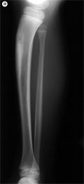
[Supracondylar process syndrome]
[INTRODUCTION - Supracondylar process is a rare bone over-growth of atavistic origin. The entity shows several similarities with the tunnel syndromes. CASE REPORT - The case reports originate from the diagnosis and the treatment of two female patients. Both of them presented with elbow region pain, weakness in finger flexion and numbness. The clinical picture, the decrease in the speed of nerve conduction in the region of the supracondylar process and radiological evidence of the process over-growth made the proper diagnosis possible in both cases. Due to the failure of conservative therapy both patients underwent surgery. Careful surgical removal of the process and neurolysis resulted in cessation of symptoms. CONCLUSION - Use of atypical radiographic positioning and flouroscopy might be required to achieve a diagnosis. In patients who do not respond to physiotherapy and local steroid injections, surgical removal of the supracondylar process is indicated which prevents further arterial and nerve degeneration.]
Hungarian Radiology
MARCH 22, 2008
Hungarian Radiology
MARCH 22, 2008

Hungarian Radiology
MARCH 22, 2008

Hungarian Radiology
MARCH 22, 2008

[Gynecological malignancies: review of the radiological diagnostics and image-guided therapy - Onco Update 2008]
[The recent results of diagnostical imaging of gynecological tumours and the actual place of interventional radiological methods are discussed. Systematical reviews of articles published during the last year (2007) have been availed to discuss: cervical cancer, endometrial cancer, ovarian cancer, general and special imaging of the female pelvis, different uterine fibroid ablation methods (embolisation and high-intensity focused ultrasound [HIFU]). Experience of gynecological tumour imaging is growing rapidly, therefore, even the current examination algorithm is changing continuously. New diagnostic and therapeutic modalities are making their way into the daily routine. Some examinations become obsolete during the course of time and thus their further application should be avoided. In the meanwhile, some modalities prove their worth and become indespensable during the investigation of a given pathology.]
Hungarian Radiology
MARCH 22, 2008

Hungarian Radiology
MARCH 22, 2008

Hungarian Radiology
MARCH 22, 2008

Hungarian Radiology
MARCH 22, 2008

Hungarian Radiology
MARCH 22, 2008

Hungarian Radiology
MARCH 22, 2008

Hungarian Radiology
MARCH 22, 2008

Hungarian Radiology
MARCH 22, 2008

1.
Clinical Neuroscience
[Headache registry in Szeged: Experiences regarding to migraine patients]
21. MAY
2.
Clinical Neuroscience
[The new target population of stroke awareness campaign: Kindergarten students ]
21. MAY
3.
Clinical Neuroscience
Is there any difference in mortality rates of atrial fibrillation detected before or after ischemic stroke?
27. NOV
4.
Clinical Neuroscience
Factors influencing the level of stigma in Parkinson’s disease in western Turkey
27. SEP
5.
Clinical Neuroscience
[The effects of demographic and clinical factors on the severity of poststroke aphasia]
18. JUL
1.
2.
3.
4.
5.