The eLitMed.hu medical portal uses computer cookies for convenient operation. Detailed information can be found in the Cookie-policy.
Hungarian Radiology - 2006;80(05-06)
Content
Hungarian Radiology
OCTOBER 20, 2006
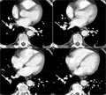
[The value of multislice computed tomography in the diagnosis of pulmonary embolism and in differential diagnostics]
[INTRODUCTION - The multislice CT (MSCT) pulmonary angiography has been used increasingly in the diagnosis of pulmonary embolism with worldwide application and nowadays it can be considered as a gold standard technique. At the author's department a single slice CT has been used from 1993 and a 10 slice CT system with 24 detector-lines was installed in 2004. The authors summarize the advantages of MSCT in the diagnosis and differential diagnosis of pulmonary embolism and its role in the follow-up of cases and the significant increase of the diagnostic safety. PATIENTS AND METHODS - 2576 chest MSCT examinations were performed between 16 February 2004 and 20 June 2005. In 261 cases pulmonary MSCT angiography was made due to suspected pulmonary embolism. In 7 cases the indication of the CT was different, but due to the findings (pulmonary embolism) this group of patients is discussed in this paper. 268 cases were evaluated retrospectively. In 12 patients, CT could not be performed due to contrast agent allergy or because of seriously impaired renal function. In 2 cases it was not possible to establish a venous access. The therapeutical result was monitored upon the request of the clinicians. The pulmonary MSCT angiography was performed in accordance with a detailed acquisition and reconstruction protocol. In the reprocessing stage the 2D (MPR) reconstruction in different directions was an important step. In addition, three-dimensional MIP was used and VRT reconstruction was also made, if needed. Bolus tracking was applied in order to achieve a good contrast phase. RESULTS - In 116 out of 268 cases pulmonary embolism was detected by pulmonary MSCT angiography. In 7 patients pulmonary embolism was not the primary diagnosis. Pathological changes could not be detected in 55 cases (20.5%) and embolism was not proven later in these patients. No false negative study was recognized. In one case, after the death of the patient the autopsy failed to prove the presence of pulmonary embolism, this represents one false positive diagnosis. In 97 patients pulmonary embolism was not detected, however other findings were found relating to the complains of the patients and thus proper therapy could be introduced. CONCLUSION - Pulmonary MSCT angiography proved to be a highly accurate method for the diagnosis and for exclusion of pulmonary embolism and also in the evaluation of its extent. MSCT can be used in monitoring the result of the therapy. In addition, MSCT gives an opportunity to detect other acute pathological conditions of the chest, thus value of the study is significant in the differential diagnosis.]


Hungarian Radiology
OCTOBER 20, 2006
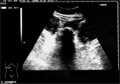
[The role of ultrasonography and X-ray examinations in the diagnosis of gallstone ileus in preoperative stage]
[INTRODUCTION - Gallstone ileus develops in elderly patients as a result of complication of cholelithiasis and causes 1-3% of mechanical ileus. Due to its variable presentation and in many cases insidious and intermittent symptoms it is difficult to establish the diagnosis which is often delayed. The mortality rate is high and early diagnosis is essential. The role of X-ray, abdominal ultrasound and recently CT has been emphasized. The role of imaging studies was evaluated in the preoperative diagnosis of gallstone ileus based on their surgically proven cases. PATIENTS AND METHODS - From 1st January 1988 to 30th June 2004 nineteen operations were performed on seventeen patients suffering from gallstone ileus. The average age of the patients was 74.2 years, male/female ratio was 4/13. Ultrasound examination was performed in all cases before the operation. Plane X-ray examination also was carried out except in two cases. CT study was not performed preoperatively. The calculi were removed through an enterotomy whole. In four cases gall bladder were also removed and the biliary fistula was closed. The disease was diagnosed on the basis of symptoms described by Rigler. If two signs from the three was present the diagnosis was established. RESULTS - 17 cases out of 19 mechanical ileus were diagnosed, in one case acute cholecystitis and in one incarcerated abdominal wall hernia was suspected before surgery. Small bowel obstruction was found in all cases (jejunum in six cases, ileum in 13). In five cases multiple calculi were seen in the bowels. Two patients had to undergo surgery twice because of gallstone ileus. It is noted that in eight cases (42.1% of total operations) gallstone ileus was diagnosed before the operation. In seven cases ultrasound played a crucial role in establishing diagnosis. In one case gas was detected in the biliary tract and in six, gallstone was directly seen in the small bowel with ultrasound. CONCLUSIONS - In the preoperative diagnosis of gallstone ileus more and more is expected from appropriate radiological examinations. If diagnosis is primarily based on the physical examination, an illusion of clinical improvement can be created, and the delay of surgical treatment can lead to decompensation of elderly patients with increased mortality rate. A thorough radiological examination can show the stone in the bowel lumen, and observation of the indirect signs together with clinical state can considerably improve the results. The rate of correct diagnosis of gallstone ileus before sugery in our patients can be considered an average and similar to the published data in medical literature.]
Hungarian Radiology
OCTOBER 20, 2006

Hungarian Radiology
OCTOBER 20, 2006
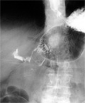
[Successful radiological diagnostics in Bouveret’s syndrome]
[INTRODUCTION - Bouveret’s syndrome I is a rare clinical entity, a special form of gallstone ileus. Based on a case study the authors describe the clinical presentation, the complications and diagnostic work up of the Bouveret’s syndrome I. CASE REPORT - A 75-year-old female patient with repeated vomiting and haematemesis was examined. Known gallstones and obstructive jaundice was noted in the case history. Urgent gastroscopy was performed at admission, which proved haemorrhagic esophagitis as the cause of the haematemesis. A gallstone was found by endoscopy distal to the pyloric region obstructing the bowel lumen. Radiological examinations proved the presence of the stone exactly at the localization that was given. Surgery confirmed the diagnosis. CONCLUSION - Bouveret’s syndrome I should be considered in patients with repeated and long lasting vomiting and bile stone in the case history. Conventional X-ray may be sufficient to establish the diagnosis, however further imaging studies are needed to clarify exact anatomical situation and potential complications of the disease.]
Hungarian Radiology
OCTOBER 20, 2006

Hungarian Radiology
OCTOBER 20, 2006

Hungarian Radiology
OCTOBER 20, 2006

Hungarian Radiology
OCTOBER 20, 2006

Hungarian Radiology
OCTOBER 20, 2006

1.
Clinical Neuroscience
[Headache registry in Szeged: Experiences regarding to migraine patients]
21. MAY
2.
Clinical Neuroscience
[The new target population of stroke awareness campaign: Kindergarten students ]
21. MAY
3.
Clinical Neuroscience
Is there any difference in mortality rates of atrial fibrillation detected before or after ischemic stroke?
27. NOV
4.
Clinical Neuroscience
Factors influencing the level of stigma in Parkinson’s disease in western Turkey
27. SEP
5.
Clinical Neuroscience
[The effects of demographic and clinical factors on the severity of poststroke aphasia]
18. JUL
1.
2.
3.
4.
5.