The eLitMed.hu medical portal uses computer cookies for convenient operation. Detailed information can be found in the Cookie-policy.
Hungarian Radiology - 2004;78(06)
Content
Hungarian Radiology
DECEMBER 20, 2004


Hungarian Radiology
DECEMBER 20, 2004

[Recent advances in the radiology of colon cancer - Onco Update 2004]
[The recent literature of colon cancer imaging, colonic stenting and the interventional treatment of colorectal liver metastases is overviewed. The introduction of virtual colonoscopy in the diagnosis of colon cancer drew attention in the last years and it is one of the most rapidly developing method. Several new publications was published about CT and MR colonography in the past one and half year. Nowadays, beside the double contrast barium enema and the colonoscopy (as gold standard), CT and MR colonography plays more and more important role. These methods can be applied only with multislice CT and modern MR machines equipped with appropriate softwares. Since these equipments are available only in limited number in Hungary, these methods has not been used in the daily practice. Development of interventional methods, like stenting of obstructive colon tumor and the percutaneous and intraarterial interventional treatment of colorectal metastases play more and more important role in the therapy, as indicated in the literature.]
Hungarian Radiology
DECEMBER 20, 2004
[Imaging studies of gynecological tumor staging]
[The imaging staging follows the surgical FIGO staging system in the classification of gynecological carcinomas taking into account the extent of tumor invasion and lymphnode involvement. The most important prognostic factors which influence treatment are the presence of invasive disease and lymphnode metastasis. Imaging techniques for evaluation of gynecological tumor staging are: abdominal and transvaginal ultrasound, computed tomography and magnetic resonance imaging. Transabdominal ultrasound is not a reliable staging modality for gynecological tumors. Computed tomography is useful in the advanced III and IV stages of diseases, but differentiation between stages I and II is difficult. Magnetic resonance imaging showed excellent diagnostic accuracy in determining adnex masses, in the assessment of cervical as well as endometrial and ovarian cancer extension. Magnetic resonance imaging is superior in comparison to computed tomography and ultrasound, in both early and advanced stage disease. Evaluation of lymph-node involvement using computed tomography and magnetic resonance imaging rely only size criteria, which is not reliable indicator of tumor involvement.]
Hungarian Radiology
DECEMBER 20, 2004
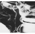
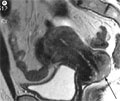
[Metastasis of the large bowel from gastric signet-ring cell carcinoma - Case report]
[INTRODUCTION - Metastatic tumor of the colon is extremely rare. It could mimic inflammatory bowel disease or ischemic colitis. CASE REPORT - A 59-year-old female underwent gastrectomy because of primary signet-ring cell carcinoma of the stomach in 1999. In November 2003 follow-up CT detected circular and segmental thickening of the wall of the transverse colon with significant targetlike contrast enchancement of the inner layers. Colonoscopy was performed but due to the significant stenosis of the transverse colon, the tumor was not properly examined. Biopsy was taken from the beginning of the stenosis, which did not prove neither specific inflammation or malignancy. Barium enema and virtual colonoscopy was performed. There was mild dilatation of the stenotic part and mucosa seemed to be intact by virtual colonoscopy. Barium enema showed significant stenosis and polypoid alteration of the mucosa in the right part of the colon. There was also an asymmetric bowel wall thickening of the flexura lienalis. Because of subileus the patient was operated on, and an extended right hemicolectomy was made. The histology of the specimen verified secondary signet-ring cell tumor of the colon. CONCLUSION - In case of multiple circular and segmental thickening of the wall of the colon, with striking thickening of the enchancing inner layer with decreased contrast enhancement in the outer layer (target pattern) on the CT, the metastasis of the large bowel should be considered.]
Hungarian Radiology
DECEMBER 20, 2004
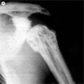
[Melorheostosis Uncommon appearances]
[INTRODUCTION - Melorheostosis is a rare but well publicised disease. About 320 cases were reported up to 1994. The incidence is estimated 0.9 cases per million. CASE REPORT - We report a 40 year-old man diagnosed with severe melorheostosis and unusual radiographic appearances. No dripping candle wax cortical thickening was present. All the long bones of the upper extremities showed extensive periosteal and endosteal hyperostosis with extension of the changes into the hands and shoulder girdle. Osteosclerosis was present at both sides of the right sacro-iliac joint. There was also hypoplasia of the scapulae, distal ulnae, carpal bones and the left thumb. Distinctive features of the clinical history were that the disease was of prenatal origin. The lower extremities were normal and in spite of extensive bone and soft tissue changes in the upper extremities this patient never experienced pain - a prominent feature of adult melorheostosis. CONCLUSION - Melorheostosis is a rare disorder, diagnosis of which can be made from clinical presentation and radiographs. In case of atypical melorheostosis, the lower extremities may be found to be normal on radiographs. The presentation may be painless.]
Hungarian Radiology
DECEMBER 20, 2004
Hungarian Radiology
DECEMBER 20, 2004

Hungarian Radiology
DECEMBER 20, 2004

Hungarian Radiology
DECEMBER 20, 2004

Hungarian Radiology
DECEMBER 20, 2004

Hungarian Radiology
DECEMBER 20, 2004
Hungarian Radiology
DECEMBER 20, 2004

Hungarian Radiology
DECEMBER 20, 2004

Hungarian Radiology
DECEMBER 20, 2004

Hungarian Radiology
DECEMBER 20, 2004

Hungarian Radiology
DECEMBER 20, 2004

1.
Clinical Neuroscience
[Headache registry in Szeged: Experiences regarding to migraine patients]
21. MAY
2.
Clinical Neuroscience
[The new target population of stroke awareness campaign: Kindergarten students ]
21. MAY
3.
Clinical Neuroscience
Is there any difference in mortality rates of atrial fibrillation detected before or after ischemic stroke?
27. NOV
4.
Clinical Neuroscience
Factors influencing the level of stigma in Parkinson’s disease in western Turkey
27. SEP
5.
Clinical Neuroscience
[The effects of demographic and clinical factors on the severity of poststroke aphasia]
18. JUL
1.
2.
Clinical Oncology
[Pancreatic cancer: ESMO Clinical Practice Guideline for diagnosis, treatment and follow-up]
29. AUG
3.
Clinical Oncology
[Pharmacovigilance landscape – Lessons from the past and opportunities for future]
29. AUG
4.
5.