The eLitMed.hu medical portal uses computer cookies for convenient operation. Detailed information can be found in the Cookie-policy.
Hungarian Radiology - 2003;77(06)
Content
[Intrauterine MR studies of the fetus]
[Ultrasound plays the primary role in the diagnostic examination to identify prenatal congenital abnormalities. In certain cases when the ultrasound is equivocal, fetal MRI can be performed in order to provide additional information to US. Based on the results of MRI, decision can be made to continue or to terminate the pregnancy. MR can be performed without premedication. No evidence of fetal risk has been reported. The new MR systems decreased the artifacts due to fetal movements, good quality studies can be done from the second trimester.]
[Comparison of different gastrointestinal contrast materials for MR examination, an experimental model]
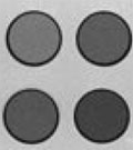
[PURPOSE - Evaluation of different gastrointestinal endoluminal contrast materials by using an experimental model. MATERIALS AND METHODS - Authors constructed a plastic container holding six plastic cups, thus making possible to evaluate different compounds and concentrations, simultaneously. The signal intensity of more than 15 different materials (commonly used contrast materials, fruit juices, cocoa, iron containing solutions) was measured by T1 and T2 weighted spin echo sequences in a 1T MR unit. The results were compared in tables and demonstrated by figures. RESULTS - The plastic container and cups made it possible to evaluate the contrast materials by MR examination. The fruit juices containing metallic components had high signal intensity on T1 weighted images, while on the T2 weighted images showed moderate to high signal intensity except the rosehip syrup and a special blackcurrant extract, which were of low signal intensity. Cocoa drink had low to moderate signal intensity on both the T1 and T2 weighted images. The signal intensity of the iron(III)-desferrioxamin solution increased on the T1 weighted images and decreased on the T2 weighted images in direct proportion to its iron concentration. CONCLUSION - The described in vitro model is an appropriate and risk-free solution for selecting the proper endoluminal contrast material, its concentration, and the best measuring sequences for defining the optimal in vivo MR bowel examination protocol. On the base of the experimental results rosehip syrup, blackcurrant extract, iron(III)- desferrioxamin and cocoa drink were selected for further in vitro and in vivo examinations.]
[Navigation for image-guided procedures: a new modality and review]
[Basic principles of two types of medical navigation are discussed. In patient-based navigation (PBN) image acquisition is followed by intervention. In modality-based navigation the imaging modality is present in the operation room, and a reference system is used. Advantages and disadvantages of both navigation types are listed. The steps of an interventional procedure with a new navigation system are described and illustrated. The role and possible future trends of navigation are finally summarized.]
[Small bowel perforation due to blunt abdominal trauma in case of an inguinal hernia]
[INTRODUCTION - The injury of fixed bowel loops occurs more frequently due abdominal trauma. Authors review the CT signs of bowel injury in conjunction of the presented case. PATIENTS, METHODS - The inguinal hernia of the male patient was present for approximately 30 years prior the abdominal trauma. Due to the trauma the fixed small bowel loop became perforated. CT examination, beside using the conventional methods established the diagnosis of bowel wall perforation and the site of the perforation was localized before surgery. CONCLUSIONS - CT provied additional information compared to X-ray and US in the localization of the lesion due to the blunt abdominal trauma.]
1.
Clinical Neuroscience
[Headache registry in Szeged: Experiences regarding to migraine patients]2.
Clinical Neuroscience
[The new target population of stroke awareness campaign: Kindergarten students ]3.
Clinical Neuroscience
Is there any difference in mortality rates of atrial fibrillation detected before or after ischemic stroke?4.
Clinical Neuroscience
Factors influencing the level of stigma in Parkinson’s disease in western Turkey5.
Clinical Neuroscience
[The effects of demographic and clinical factors on the severity of poststroke aphasia]1.
2.
Clinical Oncology
[Pancreatic cancer: ESMO Clinical Practice Guideline for diagnosis, treatment and follow-up]3.
Clinical Oncology
[Pharmacovigilance landscape – Lessons from the past and opportunities for future]4.
5.


