The eLitMed.hu medical portal uses computer cookies for convenient operation. Detailed information can be found in the Cookie-policy.
Hungarian Radiology - 2003;77(04)
Content
Hungarian Radiology
AUGUST 20, 2003


Hungarian Radiology
AUGUST 20, 2003
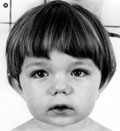
[Cheirospondyloenchondromatosis]
[The term of cheirospondyloenchondromatosis (CHE) was coined by Spranger et al. This generalised, distinctive form of enchondromatosis is characterised by mild to moderate dwarfism, short hands and feet with beaded fingers, prominent large joints and frequently mental deficiency. Major radiographic features include generalised mild platyspondyly, generalised enchondromatosis with marked involvement of hands and feet and small ilia with eroded crests and acetabular roofs. We report three patients with this rare, severe form of enchondromatosis and stress some differences between our patients and the classical description of Spranger et al.]
Hungarian Radiology
AUGUST 20, 2003

[Follow-up of infants undergoing pyeloplasty: renal ultrasound and diuresis renography changes]
[INTRODUCTION - The authors reviewed their experience after pyeloplasty in infants, according to the follow-up results of ultrasound and nuclear renography. PATIENTS AND METHODS - During the period 1988-2001 184 infants underwent pyeloplasty for ureteropelvic junction (UPJ) obstruction. Patients (n=91) with unilateral UPJ obstruction and normal contralateral kidney were included in this study. Patients ages at surgery were between 1 day-36 months. Preoperative evaluation included ultrasound examinations and diuretic renograms. Follow-up ultrasound examinations were done after 3, 6, 12 months, later yearly. Depending on the result of the ultrasound examination isotope scan were done. RESULTS - The pelviureteric obstruction were detected prenatally at 65 cases (71%). Preoperative ultrasound revealed grade 3-4 pyelocaliectasis in all patients. All patients had obstruction on preoperative renography, nine kidneys had no function, in 37 cases (41%) hydronephrotic kidneys had diminished function. Nine patients underwent nephrectomy because of non-functioning kidney. Eighty two infants underwent pyeloplasty. Grade of hydronephrosis improved in 56% of kidneys and dilatation was the same one year after pyeloplasty in 44% of kidneys. Five years after pyeloplasty 91% of kidneys, after eight years 97% of hydronephrosis improved or cured. Postoperative renography showed improvement in drainage in all preoperatively obstructed kidneys. The renal function improved only in 22% of cases at the end of the first postoperative year, and no further improvement were detected later on. CONCLUSION - Pyeloplasty in infant ages significantly cured the drainage early after surgery. The resolution of hydronephrosis is relatively slow, but after eight years dilatation cured or improved in most of the affected kidneys. Improvement of the renal function was detected only in small part of the cases.]
Hungarian Radiology
AUGUST 20, 2003
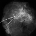
[Radial scar associated with lobular neoplasia in the breast]
[INTRODUCTION - The authors are presenting the case of a 55-year-old female patient with breast abnormalities of unclear morphology. CASE REPORT - The lesion seen in the left breast was characteristic of radial scar in which, however, numerous, but not clearly benign microcalcifications were detected. During histological examination a radial scar associated with a small lobular neoplasia was diagnosed. However, these microcalcifications were not related to the malignancy. CONCLUSION - In radial scar extensive benign microcalcifications may develop. Nevertheless we should bear in mind that in 10-30% of cases this disorder can be associated with malignancy even without mammographic signs. The final diagnosis, however, should always be made on the basis of histological examination.]
Hungarian Radiology
AUGUST 20, 2003

[Our experiences with the use of phosphor plate X-ray system and PACS]
[In this study the experiences, the advantages and disadvantages of a full digital radiology department are presented. The conventional radiology and the spot films of gastroenterologic studies are exposed on phosphor plates since 1999 at our department. Two work-stations are used for making the reports and six viewing-stations are installed at distant departments. A central server organizes the data and pictures flow and the archive system consists of magnetooptical discs in a juke-box. The conventional X-ray methods are fully integrated in the system. The number of hardcopies is dramatically decreased. The clinicians may easily access the images on the viewingstations. Possibility of teleradiology and teleconsultation is integrated in the system. The quality of the examinations is improved and became uniform. The images of different methods (CT, fluoroscopy) are stored also in digital format. The disadvantages are the high cost of installation, a new workflow and reporting habits must be initiated. A problem of one element can cause the breakdown of the whole system. The new technics, the digital world forces us to develope and define new technical standards in order to obtain uniform quality.]
Hungarian Radiology
AUGUST 20, 2003

Hungarian Radiology
AUGUST 20, 2003

Hungarian Radiology
AUGUST 20, 2003

Hungarian Radiology
AUGUST 20, 2003

Hungarian Radiology
AUGUST 20, 2003

Hungarian Radiology
AUGUST 20, 2003

Hungarian Radiology
AUGUST 20, 2003
Hungarian Radiology
AUGUST 20, 2003

Hungarian Radiology
AUGUST 20, 2003

1.
Clinical Neuroscience
[Headache registry in Szeged: Experiences regarding to migraine patients]
21. MAY
2.
Clinical Neuroscience
[The new target population of stroke awareness campaign: Kindergarten students ]
21. MAY
3.
Clinical Neuroscience
Is there any difference in mortality rates of atrial fibrillation detected before or after ischemic stroke?
27. NOV
4.
Clinical Neuroscience
Factors influencing the level of stigma in Parkinson’s disease in western Turkey
27. SEP
5.
Clinical Neuroscience
[The effects of demographic and clinical factors on the severity of poststroke aphasia]
18. JUL
1.
2.
Clinical Oncology
[Pancreatic cancer: ESMO Clinical Practice Guideline for diagnosis, treatment and follow-up]
29. AUG
3.
Clinical Oncology
[Pharmacovigilance landscape – Lessons from the past and opportunities for future]
29. AUG
4.
5.