The eLitMed.hu medical portal uses computer cookies for convenient operation. Detailed information can be found in the Cookie-policy.
Hungarian Radiology - 2002;76(01)
Content
Hungarian Radiology
FEBRUARY 20, 2002


Hungarian Radiology
FEBRUARY 20, 2002
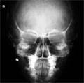
[Frequency and diagnosis of pediatric air gun injuries]
[INTRODUCTION - Air guns are frequently given to children as toys. Air guns have a pellet caliber of 0.17 or 0.22 and are propelled by compressed gas. Though they have little penetrating effect, they may cause life threatening injuries. Our purpose was to evaluate the frequency and the development of the diagnostic opportunities in children with air gun injuries during the last 30 years (1971-2000). PATIENTS AND METHODS - 52 patients (39 boys and 13 girls) were admitted to our pediatric surgery department due to of air gun injuries. The average age was 9 years (range 2 to 14 years). During the first fourteen years conventional X-ray (plain film and fluoroscopy), since 1984 ultrasonography and later (1986) CT has also been used for the diagnosis. RESULTS - In the first ten years 12 patients, in the second decade 18 patients and in the third ten years 22 patients were admitted and treated with air gun injuries. The sites of injury included upper, lower extremities (n=23), head (n=10), neck (n=5), chest (n=9) and the abdomen (n=5). The majority of patients had superficial injury and Xray plain films in different views were obtained, only. Major complication occured in 10 cases: bone fracture (n=1), soft tissue abscess (n=4) pneumothorax and hemothorax (n=4), bowel perforation (n=1). In these cases ultrasonography and/or CT was performed and they were helpful to establish the correct diagnosis. CONCLUSION - The general conception that air guns are toys, is basically wrong. The practice of placing air guns in the hands of children by their parents is very dangerous. On the basis of our results, the frequency of air gun injuries in children increased significantly in the last decade and the injuries were more serious than before (due to thew technologic modification of air gun). Ultrasonography and CT have important role in the diagnosis, but conventional X-ray remains the basic method in most of cases.]
Hungarian Radiology
FEBRUARY 20, 2002

[Current questions of quality assurance in diagnostic radiology in the light of a visit to England]
[Physical-technical aspects of quality assurance in diagnostic radiology, because of its dependence on technology are of an extraordinary importance. The intention of Hungary to join EU makes at least the decrease of our lag in this respect unavoidable. Ministerial order 31/2001 (X. 3.) EüM which already came into force requires quality assurance in diagnostic radiology explicitly. This paper starts with definition of basic concepts, then outlooks shortly the history and present international situation of quality assurance in diagnostic radiology. We review preliminaries and the present situation in Hungary, including results of the National Patient Dose Assessment Programme till now. We think that the most efficient help to the initial steps of quality assurance in diagnostic radiology in Hungary may be the appropriate adaptation of experiences of the leading countries. Therefore we review experiences of one of the authors gained during visiting three medical physics centres in England in details. The following topics are discussed: legal requirements, types and levels of measurements, organizational problems, practical evaluation of measurements (including criteria of discarding equipment), patient dosimetry, personal dosimetry, mammography research, instrumentation of the radiology departments, calibration of measuring devices, questions of the so-called type testing and radiation protection training of workers.]
Hungarian Radiology
FEBRUARY 20, 2002
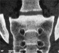
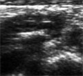
[Diagnosis of pelvic injuries]
[In most cases, the cause of the pelvic injuries are traffic accidents and falling from heights. In the seriously injuried patients the bottomline of the treatment is to establish the accurate diagnosis. The role of X-ray technicians to should be emphasized. This paper briefly summarizes the anatomical and pathological basics of the pelvic injuries, the different the types of injuries and the examination methods of choice. Beside conventional X-ray studies, CT and portable ultrasonography are also important methods with special regard in the detection of the complications associated with pelvic injuries.]
Hungarian Radiology
FEBRUARY 20, 2002
Hungarian Radiology
FEBRUARY 20, 2002

Hungarian Radiology
FEBRUARY 20, 2002

Hungarian Radiology
FEBRUARY 20, 2002

Hungarian Radiology
FEBRUARY 20, 2002

Hungarian Radiology
FEBRUARY 20, 2002

Hungarian Radiology
FEBRUARY 20, 2002

Hungarian Radiology
FEBRUARY 20, 2002

Hungarian Radiology
FEBRUARY 20, 2002

1.
Clinical Neuroscience
[Headache registry in Szeged: Experiences regarding to migraine patients]
21. MAY
2.
Clinical Neuroscience
[The new target population of stroke awareness campaign: Kindergarten students ]
21. MAY
3.
Clinical Neuroscience
Is there any difference in mortality rates of atrial fibrillation detected before or after ischemic stroke?
27. NOV
4.
Clinical Neuroscience
Factors influencing the level of stigma in Parkinson’s disease in western Turkey
27. SEP
5.
Clinical Neuroscience
[The effects of demographic and clinical factors on the severity of poststroke aphasia]
18. JUL
1.
2.
Clinical Oncology
[Pancreatic cancer: ESMO Clinical Practice Guideline for diagnosis, treatment and follow-up]
29. AUG
3.
Clinical Oncology
[Pharmacovigilance landscape – Lessons from the past and opportunities for future]
29. AUG
4.
5.