The eLitMed.hu medical portal uses computer cookies for convenient operation. Detailed information can be found in the Cookie-policy.
Clinical Neuroscience - 2017;70(01-02)
Content
Clinical Neuroscience
JANUARY 20, 2017
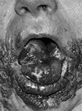
Combination of severe facial and cervical vascular malformation with obstructive sleep apnea syndrome: diagnostic and therapeutic approaches
The combination of obstructive sleep apnea syndrome and vascular malformation within the head and neck region is a rare condition, and interestingly, only a few cases have recently been published. Propagation of the vascular mass to the larynx and pharynx can cause breathing and swallowing difficulties. Due to these sypmtoms, examination and initiation of appropriate therapy for such patients are indeed challenging. We reviewed the literature available and present our case of a 64 year old woman emphasizing the complaints of sleep apnea syndrome and vascular malformation of the face and neck region. Polygraphic examination detected severe obstructive sleep apnea syndrome. The MR examination of the neck revealed extensive vascular mass narrowing the pharyngo-laryngeal region, thereby causing temporal bone destruction on the right side with intracranial propagation. ENT examination demonstrated significant narrowing of the pharyngeal lumen and the laryngeal aditus caused by multiple hemangiomas. CPAP titration showed the minimalization of the apnea-hypopnea index on the effective pressure level. Regular CPAP usage resulted in diminishing a majority of the patient’s complaints. Our examination clearly demonstrates, obstructive sleep apnea syndrome coupled with significantly obstructing vascular malformation in the head and neck region can be effectively treated safely with a CPAP device, if surgical therapy is not possible. We summarized our findings and the data available in the literature to set up recommendations for the appropriate examination and therapy (including mask fit, etc.) of vascular malformations and hemangiomas causing pharyngo-laryngeal obstruction.


Clinical Neuroscience
JANUARY 20, 2017
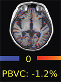
[Magnetic resonance imaging in the course of alemtuzumab and teriflunomide therapy]
[Our work aimed to review the published results of magnetic resonance imaging (MRI) obtained in the course of alemtuzumab and teriflunomide therapy in multiplex sclerosis. In multiplex sclerosis MRI sensitively detects subclinical pathological processes, which do not manifest clinically in the early course of the disease, however have substantial significance from the viewpoint of the long-term disease prognosis. MRI has an increasingly important role in the early monitoring of the therapeutic efficacy. In the last 15 years several clinical trials have been conducted with alemtuzumab and teriflunomide in multiple sclerosis providing evidence about the favourable clinical effect of these drugs. MRI images were acquired in these trials as well, and the results published recently in the scientific literature. These MRI results denote the suppression of the disease activity and the neurodegenerative processes, which may imply a favourable effect on the long-term prognosis of the disease. ]
Clinical Neuroscience
JANUARY 20, 2017
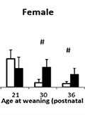
The timing of weaning alters the vulnerability to stress-induced gastric erosion in adult rats
Background - Weaning is an important period of life and its timing may influence the resilence for later stress. One of the most important stress-related disorder is gastric ulceration. Purpose and methods - Therefore we aimed to investigate the sensitivity of gastric mucosa to cold (at 16°C) water immersion stress (WIS for 3h) in adult (75-day-old) female and male rats after weaning them at different timepoints (at 17, 21, 30, 36 or 42 postnatal days). The connection with stress was studied by comparing control groups to those underwent WIS at the time of weaning and measuring corticosterone levels at the time of collecting the stomach samples. Results - The timing of weaning has strong impact on all studied parameters. Stress-induced erosion development was the smallest in rats weaned at 36-day independently from preconditioning with WIS at weaning, or sex, despite a clear sex-effect on blood corticosterone levels and body weight. WIS at weaning influenced only the body weight in adult rats weaned at 30-day, being higher in stressed than in control groups. There was no clear overall correlation between erosion area and blood corticosterone measures. Conclusions - Taken together our results confirm that the timing of weaning has long-lasting impact on the resiliance of gastric mucosa to ulcerogenic stressful events. In rats the postnatal day 30-36 seems to be optimal for weaning in both sexes as both earlier and later weaning increased vulnerability. Females seems to be more vulnerable to the effect of weaning than males.
Clinical Neuroscience
JANUARY 20, 2017

The evaluation of the relationship between risk factors and prognosis in intracerebral hemorrhage patients
Objective - Patients were assessed in terms of risk factors, hematoma size and localization, the effects of spontaneous intracerebral hemorrhage (ICH) on mortality and morbidity, and post-stroke depression. Materials and methods - The present study evaluated the demographic data, risk factors, and neurological examinations of 216 ICH patients. The diagnosis, volume, localization, and ventricular extension of the hematomas were determined using computed tomography scans. The mortality rate through the first 30 days was evaluated using ICH score and ICH grading scale. The Modified Rankin Scale (mRS) was used to determine the dependency status and functional recovery of each patient, and the Hamilton Depression Rating Scale was administered to assess the psychosocial status of each patient. Results - The mean age of the patients was 65.3±14.5 years. The most common locations of the ICH lesions were as follows: lobar (28.3%), thalamus (26.4%), basal ganglia (24.0%), cerebellum (13.9%), and brainstem (7.4%). The average hematoma volume was 15.8±23.8 cm3; a ventricular extension of the hemorrhage developed in 34.4% of the patients, a midline shift in 28.7%, and perihematomal edema, as the most frequently occurring complication, in 27.8%. Over the 6-month follow-up period, 57.9% of patients showed a poor prognosis (mRS: ≥3), while 42.1% showed a good prognosis (mRS: <3). The mortality rate over the first 30 days was significantly higher in patients with a low Glasgow Coma Scale (GCS) score at admission, a large hematoma volume, and ventricular extension of the hemorrhage (p=0.0001). In the poor prognosis group, the presence of moderate depression (39.13%) was significantly higher than in the good prognosis group (p=0.0001). Conclusion - Determination and evaluation of the factors that could influence the prognosis and mortality of patients with ICH is crucial for the achievement of more effective patient management and improved quality of life.
Clinical Neuroscience
JANUARY 20, 2017
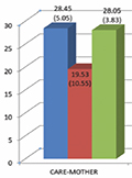
Symptom profiles and parental bonding in homicidal versus non-violent male schizophrenia patients
Objective - To compare the intensity and the profile of psychotic symptoms and the characteristics of parental bonding of male schizophrenia patients with a history of homicide and those without a history of violent behaviour. Clinical question - We hypothesized more intense psychotic symptoms, especially positive symptoms as signs of a more severe psychopathology in the background of homicidal behaviour. We also hypothesized a more negatively perceived pattern (less Care more Overprotection) of parental bonding in the case of homicidal schizophrenia patients than in non-violent patients and non-violent healthy controls. Method and subjects - Symptom severity and symptom profiles were assessed with the Positive and Negative Syndrome Scale in a group of male schizophrenia patients (n=22) with the history of committed or attempted homicide, and another group (n=19) of male schizophrenia patients without a history of violent behaviour. Care- and Overprotection were assessed using the Parental Bonding Instrument (PBI) in a third group of non-violent healthy controls (n=20), too. Results - Positive, negative and general psychopathology symptoms in the homicidal schizophrenia group were significantly (p<0.005) more severe than in the non-violent schizophrenia group. Non-violent schizophrenia patients scored lower on Care and higher on Overprotection than violent patients and healthy controls. Homicidal schizophrenia patients showed a pattern similar to the one in the healthy control group. Conclusions - It seems imperative to register intense positive psychotic symptoms as predictive markers for later violent behaviour. In the subgroup of male homicidal schizophrenia patients negatively experienced parental bonding does not appear to be major contributing factor to later homicidal behaviour.
Clinical Neuroscience
JANUARY 20, 2017
Eating behaviors among the participants of an inpatient weight loss treatment
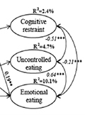
Background and purpose - Eating behaviors play a crucial role in the development and maintenance of excess weight. The aim of the study was to explore the predictors and changes in eating behaviors among overweight and obese patients. Methods - The sample of the 6-month prospective survey consisted of patients who participated in the inpatient weight loss treatment program in the Lipidological Department of the Szent Imre Hospital (baseline: N=339, 19% men; follow-up: N=175, 16% men). The mean age was 50.2 years (SD=13.47), the mean BMI was 38.6 (SD=7.58) at baseline. Measures: self-reported anthropometric data, Three-Factor Eating Questionnaire Revised 21-Items, CES-D Depression Scale. Results - According to the results of Multiple Indicators and Multiple Causes analysis, older age predicted greater cognitive restraint (b=0.12, p=0.047). Women were more prone to emotional eating than men (b=0.21, p<0.001). Higher levels of education predicted greater uncontrolled eating (b=0.16, p=0.007) and emotional eating (b=0.12, p=0.039). Depression showed a positive relationship with emotional eating (b=0.19, p=0.001), and mediated the relationship between gender and emotional eating (b=0.04, p=0.009), and BMI and emotional eating (b=0.03, p=0.015). Those whose weight loss was at least 5% showed a greater improvement in the eating behaviors than those whose weight loss was below 5% (cognitive restraint: t(168)=-4.765, p<0.001, uncontrolled eating: t(168)=-2.442, p=0.016, and emotional eating: Z=-2.011, p=0.044). Conclusions - Results reveal certain determinants of eating behaviors that enhance or obstruct successful long term weight loss and highlight the role of eating behavior changes in weight loss. These mark intervention points for the optimization of results achievable by weight loss treatments.
Clinical Neuroscience
JANUARY 20, 2017

Patient with a spontaneously evolving carotid cavernous fistula in the emergency department
Background - Approximately 2% of patients admitted to the emergency department present with headache, which is often associated with vomiting, ocular pain, and earache. In rare cases, the presence of an abnormal communication between a cavernous sinus and the carotid arterial system that creates a carotid cavernous fistula is the main cause of these symptoms. Case presentation - A 32-year-old woman presented at the emergency department with unilateral headache associated with earache on the same side, and pulsating tinnitus. On examination, we observed unusual appearance of our patient (small stature, unusually visible skin, lobeless ears). In the first 5 hours of our observation no neurological symptoms had been present, but after a severe vomiting, exophthalmos, subconjunctival suffusion and moderate ptosis developed. First, regarding the initial general symptoms, otorhinolaryngologist assessed the patient, and did not find any abnormality. Further, we ordered computed tomography and consulted a neurologist. Despite of the negative results we continued the observation because her symptoms did not improve. After appearance of neurological symp-toms, carotid cavernous fistula was suspected. Magnetic resonance imaging and ophthalmologist consultation verified the diagnosis. For therapy, she was transferred to interventional neuroradiology. Because of the unusual appearance and carotic cavernous fistula, we ordered genetic examination. This indicated the presence of Ehlers-Danlos syndrome type IV in the background. The first major manifestation of the syndrome was observed at our department. Conclusions - Carotid cavernous fistula is an uncommon diagnosis in the emergency department; however, the early recognition of symptoms and early treatment can prevent further consequences of this potentially severe condition.
Clinical Neuroscience
JANUARY 20, 2017
[Thrombolysis in case of ischemic stroke caused by aortic dissection]
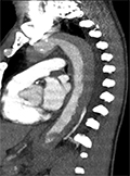
[Seldom, an acute aortic dissection can be the etiology of an acute ischemic stroke. The aortic dissection typically presents with severe chest pain, but in pain-free dissection, which ranges between 5-15% of the case, the neurological symptoms can obscure the sypmtos of the dissection. By the statistical data, there are 15-20 similar cases in Hungary in a year. In this study we present the case history of an acute ischemic stroke caused by aortic dissection, which is the first hungarian publication in this topic. A 59-year-old man was addmitted with right-gaze-deviation, acute left-sided weakness, left central facial palsy and dysarthric speech. An acute right side ischemic stroke was diagnosed by physical examination without syptoms of acute aortic dissection. Because, according to the protocol it was not contraindicated, a systemic intravenous thrombolysis was performed. The neurological sypmtoms disappeared and there were no complication or hypodensity on the brain computed tomography (CT). 36 hours after the thrombolysis, the patient become restlessness and hypoxic with back pain, without neurological abnormality. A chest CT was performed because of the suspition of the aortic dissection, and a Stanford-A type dissection was verified. After the acute aortic arch reconstruction the patient died, but there was no bleeding complication at the dissection site caused by the thrombolysis. This case report draws attention to the fact that aortic dissection can cause acute ischemic stroke. Although it is difficult to prove it retrospectively, we think the aortic dissection, without causing any symptoms or complain, had already been present before the stroke. In our opinion both the history of our patient and literature reviews confirms that in acute stroke the thrombolysis had no complication effect on the aortic dissection but ceased the neurological symptoms. If the dissection had been diagnosed before the thrombolysis, the aortic arch reconstruction would have been the first step of the treatment, without thrombolysis. ]
1.
Clinical Neuroscience
[Headache registry in Szeged: Experiences regarding to migraine patients]
21. MAY
2.
Clinical Neuroscience
[The new target population of stroke awareness campaign: Kindergarten students ]
21. MAY
3.
Clinical Neuroscience
Is there any difference in mortality rates of atrial fibrillation detected before or after ischemic stroke?
27. NOV
4.
Clinical Neuroscience
Factors influencing the level of stigma in Parkinson’s disease in western Turkey
27. SEP
5.
Clinical Neuroscience
[The effects of demographic and clinical factors on the severity of poststroke aphasia]
18. JUL
1.
2.
3.
4.
5.