The eLitMed.hu medical portal uses computer cookies for convenient operation. Detailed information can be found in the Cookie-policy.
Clinical Neuroscience - 2016;69(07-08)
Content
Clinical Neuroscience
JULY 30, 2016

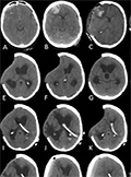
[Meningioma and pregnancy]
[Meningiomas are one of the most frequent primary intracranial tumours, representing one-third of all intracranialneoplasms. The vast majority of meningiomas are histologically benign, but recurrence and progression is quite frequent. They occur usually between the 6th and 7th decade, the female/male ratio is 3:2. Although rare in pregnancy, when occurring, they can cause serious, life-threatening complications due to rapid growth and unfavourable localisation. There are two dominant hypothesis explaining rapid growth in pregnancy: the role of hormonal effects and hemodynamic changes. Several studies tested these theories but none provided unequivocal answer probably because the pathomechanism is complex and multifactorial. We provide an overview of the pathomechanism of meningiomas in pregnancy with emphasis on data obtained by advanced neuropathological, molecular biological, bioinformatic, imaging and epidemiological methods. A better understanding of the processes leading to meningioma development and growth in pregnancy will help us to design personalized therapy and reduce morbidity and mortality.]


Clinical Neuroscience
JULY 30, 2016
Syndrome of trephined-underestimated and poorly understood complication after decompressive craniectomy
Decompressive craniectomy (DC) is still a matter of debate, with a numerous complications as expansion of haemorrhagic contusions, external cerebral herniation, subdural hygromas, post-traumatic hydrocephalus (HC). The often overlooked “syndrome of the trephined” (ST) as a delayed complication of DC also known as sinking skin flap sy initially described in 1939.ST is characterised by the neurological changes associated with alteration of the pressure/volume relationship between intracranial pressure (ICP), volume of cerebrospinal fluid (CSF), blood, and brain tissue in patients with large bone defects. This review aims at elucidating the mechanisms responsible for the development of ST, and providing useful tips and red-flag signs for healthcare professionals involved with care of post DC patients. Symptoms identified on time could help to develop appropriate treatment strategies for this suddenly deteriorating, but possible reversible condition. Although the treatment strategy is straightforward, calling for a prompt cranioplasty, the correction of HC through CSF diversion devices might require a lengthy optimisation period. Continuous changes in the setting of the shunting systems or spinal tap might lead to dangerous swinging of the midline structures causing further neurological deterioration. Thus, finding the right balance in terms of clinical management often represents a significant challenge.
Clinical Neuroscience
JULY 30, 2016

[The role of zonisamide in the management of pediatric partial epilepsy]
[In our review we discuss the group of approved antiepileptic drugs for children in Hungary. We cite the results of the review conducted by the International League Against Epilepsy on antiepileptic drug efficacy and effectiveness as initial monotherapy for newly diagnosed epileptic seizures and syndromes in pediatric age group. 25% of pediatric epilepsy is therapy resistant, so we further need new drugs, which must be investigated according to the rules of the European Medicine Agency. The ethical dilemmas of childhood drug studies lead to the situation that the new antiepileptic drugs, approved as monotherapy in adult epilepsies, are in the majority just in add-on regimen tested in pediatric patients. As clinicians we appreciate open label extension safety studies. An old-new antiepileptic drug in Europe is zonisamide. Though it was approved for first line monotherapy in pediatric and adult patients with partial and generalised epilepsy in 1989 in Japan, the European Medicine Agency licensed its use as adjunctive therapy in children aged 6 years or older with partial seizures (with or without secondary generalisation) just in 2013. The results of the openlabel extension study appeared in 2014. The mean dose received was 7.5 mg/kg/day. During the open label phase 11% of the patients achieved seizure freedom and it was maintained throughout the study. The drug was generally well tolerated. The most frequently reported treatment-related adverse events were decreased weight (6%), decreased appetite (4%), and headache (2%). No new or unexpected side effects emerged. In conclusion oral zonisamide as adjunctive therapy in pediatric patients aged 6-17 years with partial seizures demonstrated an acceptable safety and tolerability profile and efficacy over a period at least 1 year. So it is a good treatment option in this population.]
Clinical Neuroscience
JULY 30, 2016
[Angioneuritic edema in ischaemic stroke patients treated with rt-PA]
[Data of our 254 patients who were treated with rt-PA between 1st of Jan, 2011 and 31st of Dec, 2014 were processed. We focused on angioneurotic oedema as allergic complication of thrombolysis which caused life threatening respiratory obstruction in two cases. We describe these two patients’ history. Out of 254 patients six (2.3%) suffered angioneurotic edema caused respiratory obstruction in two (0.90%) cases. This occurrence is approximately 1.3-5.1% in literature. Five, out of six patients who suffered from angioneurotic oedema, had been treated with ACE inhibitors or ARB before. The role of ACE inhibitors is known in metabolism of bradykinin cascade. Plasmin which present during thrombolysis, precipitates biochemical mechanisms of this potential life threatening complication. Therefore rt-PA alone can be the cause of angioedema, but it can be more frequent together with ACE inhibitors therapy.]
Clinical Neuroscience
JULY 30, 2016
[Transthyretin familial amyloid polyneuropathy - three Hungarian cases with rare mutations (His88Arg and Phe33Leu)]
[Introduction - Transthyretin familial amyloid polyneuropathy is a rare autosomal dominant progressive systemic disesase of adults caused by endoneural amyloid deposition due to point mutations of the transthyretin gene. It is the most severe form among hereditary polyneuropathies, being fatal within 10 years if left untreated. The disease is underdiagnosed, the late onset forms (above the age of 50) being probably more widespread than previously thought. Early diagnosis is essential as the early introduction of causal therapy (tafamidis) slows progression and prolongs survival. Patients - We report here three non-related Hungarian cases of transthyretin familial amyloid polyneuropathy with non- Val30Met mutations (His88Arg in two cases, Phe33Leu in one case). They were all characterized by late-onset, progressive, length-dependent, axonal, sensorimotor polyneuropathy and the simultaneous presentation of severe restrictive cardiomyopathy. In all three cases, clinical and electrophysiological signs of myopathy were also present, suggesting the involvement of skeletal muscles as well. In two cases, high resolution ultrasound of the peripheral nerves was also performed, which showed segmental structural alterations (change or loss of fascicular structure) and some increase of echogenicity of the interfascicular epineurium, without substantial enlargement of the nerves. Conclusion - In Hungary, mainly the rare, non-Val30Met mutation forms of transthyretin familial amyloid polyneuropathy are encountered, as in our cases. As opposed to the Val30Met forms, these mutations are characterized by late onset and simultaneous presentation of severe cardiomyopathy. Our report highlights the importance of considering transthyretin familial amyloid polyneuropathy in the differential diagnosis of late-onset, progressive, axonal polyneuropathies of unknown etiology, particularly if associated with cardiac disease.]
Clinical Neuroscience
JULY 30, 2016

[The effect of anesthesia on cognitive functions]
[Aim of the study - General anesthetics, arterial hypotension and hypoxia developing during anesthesia may result in impaired memory and a decline in other abilities (such as attention, concentration, linguistic and writing abilities). Our aim was to detect changes in cognitive functions due to surgery and anesthesia with controlled arterial hypotension. Materials and methods - We studied combined and intravenous anesthesia detecting pre-and postoperative cognitive functions, intraoperative haemodynamic parameters, demographic data, other data of case history and surgical data. The Montreal Cognitive Assessment test was applied in the randomized, prospective study. The preoperative data served as basis for comparison. The second test was performed following surgery when patients were fully awake. Both groups included 30 patients. Results and conclusion - After both narcosis methods (postoperative second hour) cognitive functions were significantly deteriorated (p<0.05). Median MoCA before sevoflurane anesthesia was 24 points (interquartile range: 22-25), postoperative value was 20 (19-21) (p<0.05). Median MoCA before propofol anesthesia was 24 points (23-26), postoperative value was 20 (18-22) (p<0.01). Intraoperative arterial blood pressure, pulse rate and oxygen saturation values did not correlate with worsening of cognitive function (Pearson correlation coefficient values between -0.19 and 0.42). Execution is influenced by age (negative correlation) and education (positive correlation).]
Clinical Neuroscience
JULY 30, 2016

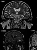
Gray matter atrophy in presymptomatic Huntington’s patients
Background - Huntington’s disease is a progressive disease in which neurodegeneration is on-going from the early presymptomatic phase. Development of sensitive biomarkers in this presymptomatic stage that are able to monitor the disease progression and test the efficacy of putative neuroprotective treatments are essential. Methods - Seven presymptomatic Huntington mutation carriers and ten age-matched healthy controls were recruited. Six of the patients participated in a 24 months longitudinal study having MRI scans 12 and 24 months after the baseline measurements. High resolution T1 weighted images were carried out and voxel based morphometry was used to analyse the data. Apart of group differences, correlation of CAG repeat number with focal cortical thickness and with global gray matter volume was calculated. Results - Focal cortical atrophy was found bilaterally in the superior temporal sulcus and in the left middle frontal gyrus in presymptomatic Huntington patients in whom no sign of cognitive or motor deterioration was detected. Global gray matter atrophy (p<0.048) and decreased total brain volume was found. The number of CAG triplets showed no correlation with the focal gray matter atrophy and total brain volume. Strong correlation between the CAG repeat number and global gray matter volume was found (p<0.016). Conclusion - Cortical atrophy is apparent in the early, presymptomatic stage of the disease. With further validation in large patient sample atrophy measure could be biomarker of disease progression and putatively of neurodegeneration.
Clinical Neuroscience
JULY 30, 2016

Burning mouth syndrome: Evaluation of clinical and laboratory findings
Background and purpose - Burning mouth syndrome is a chronic and persistent painful condition characterized by burning sensation in the oral mucosa. We investigated the etiological factors of patients presented with the history of burning in the mouth who admitted our outpatient clinics over the 8-years period and who had no underlying identifiable local factors. We also tried to determine their demographic and clinical characteristics. Our aim was to investigate the association between burning mouth and psychiatric disorders such as depression and anxiety, chronic diseases like diabetes mellitus (DM) and other laboratory studies in patients complaining of solely burning in the mouth. Methods - The study included patients with the history of burning in mouth who presented in our outpatient clinic between 2005 and 2012. They were evaluated by a neurologist, a psychiatrist, an internist, and a dentist. Complete blood counts, biochemical analysis and cranial magnetic resonance imaging (MRI) were performed for all patients. Results - A total of 26 (22 (84%) females, 4 (15%) males; mean age 55.9 years) patients were enrolled in this study. Five (19.2%) of the patients had depression, 2 (7.7%) had anxiety disorder, 2 (7.7%) had diabetes mellitus, 8 (30%) had B12 vitamin deficiency, 3 (11.5%) had decreased ferritin levels in blood, and 1 (3.8%) had folic acid deficiency. Cranial MRI of all patients were normal. Nine patients (34.6%) had no etiological causes. Conclusion - A multidisciplinary approach in the management of burning mouth and establishment of common criteria for the diagnosis would provide insight into the underlying pathophysiological mechanism.
Clinical Neuroscience
JULY 30, 2016
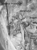
Association of anterior thoracic meningocele and azygos lobe of the lung
Here we report an anterior thoracic meningocele case. Twoyears- old female patient was presented with kyphosis. Azygos lobe of the lung was also demonstrated during radiological studies. Posterolateral thoracotomy incision and extralpeural approach was performed for excision of the anterior meningocele to untether the cord. Although both anomalies are related to faulty embryogenesis and it is well known that faulty embryogenesis may also reveal coexisting abnormalities, we could not speculate a common mechanism for anterior thoracic meningocele and azygos lobe of the lung association.
Clinical Neuroscience
JULY 30, 2016
Four cases of GABAB receptor encephalitis
GABAB receptor (gamma-aminobutyric acid type B receptors - GABABR) encephalitis is a rare manifestation of autoimmune encephalitides. We report four cases - including the first two Hungarian patients - with some peculiar features. One patient developed subacute disorientation and almost complete loss of short-term memory, but no epilepsy. Without immunotherapy, his memory spontaneously improved up to mild cognitive impairment in six weeks. GABABR antibodies persisted in his serum, and 18 months later, FDG-PET detected abnormal mediastinal lymph nodes and small cell lung cancer (SCLC). Another patient had persistently decreased sodium content in the peripheral blood. In those three patients who died, CSF was abnormal, but CSF was not pathological in the patient, who spontaneously improved. Brain MRI indicated signal intensity changes in the medial temporal areas in three cases. SCLC was found in three patients. Only the patient, who spontaneously improved, survived for more than 24 months. In summary, our cases show that (i) GABABR encephalitis may develop without epilepsy; (ii) the severe short-term memory loss can spontaneously improve; (iii) persistent hyponatremia can be present in the blood; (iv) the patient with benign course without epilepsy and CSF abnormality survived; (v) spontaneously remitting encephalitis can precede SCLC by 1.5 year, which emphasizes that repeated search for cancer is of paramount importance even in cases with spontaneous improvement.
1.
Clinical Neuroscience
[Headache registry in Szeged: Experiences regarding to migraine patients]
21. MAY
2.
Clinical Neuroscience
[The new target population of stroke awareness campaign: Kindergarten students ]
21. MAY
3.
Clinical Neuroscience
Is there any difference in mortality rates of atrial fibrillation detected before or after ischemic stroke?
27. NOV
4.
Clinical Neuroscience
Factors influencing the level of stigma in Parkinson’s disease in western Turkey
27. SEP
5.
Clinical Neuroscience
[The effects of demographic and clinical factors on the severity of poststroke aphasia]
18. JUL
1.
2.
Clinical Oncology
[Pancreatic cancer: ESMO Clinical Practice Guideline for diagnosis, treatment and follow-up]
29. AUG
3.
Clinical Oncology
[Pharmacovigilance landscape – Lessons from the past and opportunities for future]
29. AUG
4.
5.