The eLitMed.hu medical portal uses computer cookies for convenient operation. Detailed information can be found in the Cookie-policy.
Clinical Neuroscience - 2009;62(09-10)
Content
Clinical Neuroscience
OCTOBER 20, 2009
[Sexological problems in neurological disorders: neurosexology]
[The author has examined this complex subject-matter as he has not found any publications dealing with the interconnection between neurology and sexuality in the Hungarian literature available to him. Healthy sexual behavior determines the individual’s quality of life. This, however requires a coordinated, complex functioning bound to very complex structures and their unimpaired functions: peripheral receptor→ peripheral nerve→radix→spinal cord→ definite, functionally interrelated structures of the brain (prae-optic areas, hypothalamus, amygdala, limbic system and the cerebral cortex, mainly the orbitofrontal area). The functioning of these structures and the healthy sexuality are also influenced by steroid hormones, neurochemical regulations, neurotransmitters, the monoamin system, opioids, GABA, neuroendocrine hormones (oxytocin, prolactin, gonadotrop realising hormone). The author deals in detail with the impairment for some reason of neurological structures participating in sexuality, which may lead to sexual dysfunctions. ]


Clinical Neuroscience
OCTOBER 20, 2009

[Pompe’s disease - Part II - Treatment strategies and enzyme replacement]
[Pompe’s disease is an ultra-orphan disease caused by the deficiency of lysosomal alpha-glucosidase. At present, it is the only inherited muscle disorder, which can be treated by replacement of the enzyme. Three international randomized trials examined the clinical efficacy of enzyme replacement therapy (ERT) in infantile and late-onset diseases. ERT reduced the risk of death, respiratory support, invasive ventilation and improved cardiomyopathy. Respiration, muscle function and quality of life were improved in both infantile and late-onset diseases. These randomized and pilot trials also proved the safety of the treatment. At present it is not clear if antibodies induced by ERT result in decreased efficacy. In this review, we also discuss our experiences obtained by the treatment of three patients, and review the spectrum of supportive and experimental treatment strategies.]
Clinical Neuroscience
OCTOBER 20, 2009
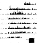
[Actigraphy: A valuable diagnostic tool or a luxury investigation? (Neuropsychiatric aspects)]
[Aim - Despite of the continuing interest in actigraphy it has a relatively low impact on the everyday medical routine. Accordingly, we set out to review the current state and to recommend relevant further indications for its application, especially in neuropsychiatry. Method - We surveyed the areas of its use and then reviewed the literature, with special regard to its advantages and methodological limitations. Adding to the results we enlarged the results with our own personal experience. Results - The limitations of the method may be decreased by methodological manipulations (a simple rational data reduction is recommended). Actigraphy appears to be a useful tool in the diagnosis of illnesses, syndromes or disorders influencing the vigilance level, the motor performance or the energetic balance of the organism. We constructed a list of the most relevant fields of indication of actigraphy in diagnostics, illustrated by anecdotal actigraphic case records. Conclusion - Further methodological considerations are needed for the successful evaluation of accelerometry. Targeted basic epidemiological studies in the healthy population and in patient groups should solve various open questions in order to make full use of the advantages of actigraphy in the everyday clinical routine.]
Clinical Neuroscience
OCTOBER 20, 2009
[Interhemispheric propagation of seizures in mesial temporal lobe epilepsy]
[Objectives - To investigate interhemispheric propagation of mesial temporal lobe epilepsy seizures in patients undergoing long-term video-EEG monitoring with combined scalp and foramen ovale electrodes. Aim of the study - To reveal possible interhemispheric propagation patterns in mesial temporal lobe epilepsy, to improve presurgical evaluation of temporal epileptic patients. Methods - Sixty-five seizures from 20 patients were analyzed. We defined two contralateral seizure propagation patterns: Type I for those seizures that spread to the contralateral foramen ovale electrodes earlier than to the contralateral scalp electrodes, and type II for the opposite. Participants - Twenty drug resistant epileptic patients were investigated in frame of their presurgical evaluation. Results - The majority of seizures (80%) were classified as type I. Inter-foramen ovale electrode propagation time was significantly shorter for type I compared to type II seizures. Ninety percent of patients had either type I or type II seizures only. Patients with type I seizures significantly more often had mesiotemporal structural alterations evident on magnetic resonance imaging scans, and became more often seizure-free after surgery compared to patients with type II seizures whose surgical outcome was less favorable or surgery could not be indicated because of independent bilateral ictal seizure-onset. Conclusions - The two types of contralateral propagation patterns we are describing seem to represent two subtypes of mesial temporal lobe epilepsy with different morphological and prognostic features. The predominance of type I over type II seizures together with shorter propagation times for type I seizures indicate a role of a more direct and dominant interhemispheric pathway in mesial temporal lobe epilepsy.]
Clinical Neuroscience
OCTOBER 20, 2009
[The transcription of the amyloid precursor protein and tryptophan 2,3-dioxygenase genes are increased by aging in the rat brain]
[Aging itself is considered as a major risk factor of dementia. The prevalence of the Alzheimer’s disease (AD) is increasing exponentially after the age of 65 and doubles every 5 years. The major aim of our present research was to examine the effect of aging on the transcription of certain genes associated with neurodegenerative disorders in the rat brain. The influence of the vasopressin (VP) hormone was also examined in the same experimental paradigm. Age dependent transcriptional changes of the following four genes were examined in the cerebral cortex: the first was the gene of the amyloid precursor protein (APP) which is abnormally cleaved to toxic beta-amyloid fragments. These aggregated peptides are the major components of the senile plaques in the AD brain. The second one was the mitogen-activated protein kinase (MAPK1) gene. The MAPK is involved in the abnormal hyperphosphorylation of the tau-protein which results in aggregated neurofibrillary tangles. The beta-actin gene was the third one. The protein product of this gene is considered to be involved in synaptogenesis, neuronal plasticity and clinical conditions like depression and AD. The last one was the gene of the tryptophan 2,3-dioxygenase (TDO2) enzyme. The activity of this enzyme is considered as a rate limiting factor in the metabolism of the neuro-immune modulator quinolinic acid (QUIN). The transciptional activity of young (2.5 months) and aged (13 months) Brattleboro rats with or without VP expression were compared by means of real time PCR technique. The cortical transciptional activity of the APP and TDO2 genes were increased in the aged animals as compared with the activity of the young ones, and this effect was independent on the presence of the VP. Our results indicate the importance of certain age dependent transcriptional changes might influence the mechanism of AD and other neurodegenerative disorders.]
Clinical Neuroscience
OCTOBER 20, 2009

Clinical Neuroscience
OCTOBER 20, 2009

[Account on the scientific meeting of the Környey Society in 2009]
[Account on the scientific meeting of the Környey Society in 2009 2009;62(09-10)]
Clinical Neuroscience
OCTOBER 20, 2009

Clinical Neuroscience
OCTOBER 20, 2009
Clinical Neuroscience
OCTOBER 20, 2009

1.
Clinical Neuroscience
[Headache registry in Szeged: Experiences regarding to migraine patients]
21. MAY
2.
Clinical Neuroscience
[The new target population of stroke awareness campaign: Kindergarten students ]
21. MAY
3.
Clinical Neuroscience
Is there any difference in mortality rates of atrial fibrillation detected before or after ischemic stroke?
27. NOV
4.
Clinical Neuroscience
Factors influencing the level of stigma in Parkinson’s disease in western Turkey
27. SEP
5.
Clinical Neuroscience
[The effects of demographic and clinical factors on the severity of poststroke aphasia]
18. JUL
1.
2.
3.
4.
5.