The eLitMed.hu medical portal uses computer cookies for convenient operation. Detailed information can be found in the Cookie-policy.
Clinical Neuroscience - 2008;61(07-08)
Content
Clinical Neuroscience
JULY 30, 2008
[Evolution of human brain and intelligence]
[The biological evolution, including human evolution is mainly driven by environmental changes. Accidental genetic modifications and their innovative results make the successful adaptation possible. As we know the human evolution started 7-8 million years ago in the African savannah, where upright position and bipedalism were significantly advantageous. The main drive of improving manual actions and tool making could be to obtain more food. Our ancestor got more meat due to more successful hunting, resulting in more calory intake, more protein and essential fatty acid in the meal. The nervous system uses disproportionally high level of energy, so better quality of food was a basic condition for the evolution of huge human brain. The size of human brain was tripled during 3.5 million years, it increased from the average of 450 cm3 of Australopithecinae to the average of 1350 cm3 of Homo sapiens. A genetic change in the system controlling gene expression could happen about 200 000 years ago, which influenced the development of nervous system, the sensorimotor function and learning ability for motor processes. The appearence and stabilisation of FOXP2 gene structure as feature of modern man coincided with the first presence and quick spread of Homo sapiens on the whole Earth. This genetic modification made opportunity for human language, as the basis of abrupt evolution of human intelligence. The brain region being responsible for human language is the left planum temporale, which is much larger in left hemisphere. This shows the most typical human brain asymmetry. In this case the anatomical asymmetry means a clearly defined functional asymmetry as well, where the brain hemispheres act differently. The preference in using hands, the lateralised using of tools resulted in the brain asymmetry, which is the precondition of human language and intelligence. However, it cannot be held anymore, that only humans make tools, because our closest relatives, the chimpanzees are able not only to use, but also to make tools, and they can be tought how to produce quite difficult ones. Some brain characteristics connected to human consciousness and intelligence, like brain asymmetry, the “consciousness” or “theory of mind” based on mirror neurons are surprisingly present in monkeys. Nevertheless, the human intelligence is extremly flexible and different, while the animal intelligence is specialised, producing one thing at high level. Based on recent knowledge the level of intelligence is related anatomically to the number of cortical neurons and physiologically to the speed of conductivity of neural pathways, the latter being dependent on the degree of myelinisation. The improvement of cognitive functions including language is driver by the need of more effective communication requiring less energy, the need of social dominance, the competitive advantages within smaller groups and species or against other species, which improves the opportunity for obtaining food. Better mental skills give also sexual dominance, which is beneficial for stabilising “cleverness” genes. The evolutionary history of human consciousness emphasises its adaptive survival helping nature. The evolution of language was the basic condition of conscious thinking as a qualitative change, which fundamentally differentiate us from all other creatures.]


Clinical Neuroscience
JULY 30, 2008

[BRAIN LATERALIZATION AND SEIZURE SEMIOLOGY: ICTAL CLINICAL LATERALIZING SIGNS]
[Clinical lateralizing signs are the phenomena which can unequivocally refer to the hemispheric onset of epileptic seizures. They can improve the localization of epileptogenic zone during presurgical evaluation, moreover, their presence can predict a success of surgical treatment. Primary sensory phenomena such as visual aura in one half of the field of vision or unilateral ictal somatosensory sensation always appear on the contralateral to the focus. Periictal unilateral headache, although it is an infrequent symptom, is usually an ipsilateral sign. Primary motor phenomena like epileptic clonic, tonic movements, the version of head ubiquitously appear contralateral to the epileptogenic zone. Very useful lateralization sign is the ictal hand-dystonia which lateralizes to the contralateral hemisphere in nearly 100%. The last clonus of the secondarily generalized tonic-clonic seizure lateralizes to the ipsilateral hemisphere in 85%. The fast component of ictal nystagmus appears in nearly 100% on the contralateral side of the epileptic focus. Vegetative symptoms during seizures arising from temporal lobe such as spitting, nausea, vomiting, urinary urge are typical for seizures originating from non-dominant (right) hemisphere. Ictal pallor and cold shivers are dominant hemispheric lateralization signs. Postictal unilateral nose wiping refers to the ipsilateral hemispheric focus compared to the wiping hand. Ictal or postictal aphasia refers to seizure arising from dominant hemisphere. Intelligable speech during complex partial seizures appears in non-dominant seizures. Automatism with preserved consciousness refers to the seizures of non-dominant temporal lobe.]
Clinical Neuroscience
JULY 30, 2008

[IMPORTANT AIM OF STATIN THERAPY: ISCHEMIC CARDIOVASCULAR EVENT (STROKE)]
[Statin’s treatment clearly is authorized in prevention of coronary heart disease (CHD). According to the results of many studies and meta-analyses statins can inhibit the first cerebrovascular infarct (stroke). The greater the decrease of LDL-cholesterol level the more prominent the efficacy. The effect is not so robustic compared to coronaria vessels moreover clear pleiotropic (cholesterol independent) action takes also part. It has been nowadays revealed that high dose (80 mg) atorvastatin can confine first time the development of recurrence stroke (SPARCL study), which is an important fact in the field of secondary prevention.]
Clinical Neuroscience
JULY 30, 2008

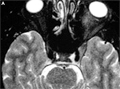
[SUCCESSFULL SURGICAL REMOVAL OF A MESENCEPHALIC CAVERNOUS ANGIOMA, WHICH WAS RESPONSIBLE FOR PROGRESSIVE NEUROLOGICAL DEFICITS]
[Cavernous angiomas comprise 5-10% of all vascular malformations in the central nervous system, occuring most frequently in the supratentorial region, and 20% of them in the brain stem. According to literature, brain stem cavernous angiomas occur most frequently in the pons (60%), and equally in the mesencephalon (20%) and in medulla oblongata. In clinical evaluation the authors describe the successful removal of a mesencephalic cavernous angioma causing progressive neurological deficits and symptoms. The authors present a case of a 51 year old female, who had developed 1 year prior to her admittance: fatigue, weakness in the right upper limb and fingers, right lower limb ataxia. One month later, her lower right limb developed sensory deficits. The first neurological exploration indicated dysarthria, moderate facial and right hemiparesis, hemihypaesthesia and ataxia. CT and MR imaging indicated multilobulated cavernomas in the mesencephalon. After conservative treatment the patient became almost symptom free, and thus neurosurgical treatment was not discussed. Later on her symptoms fluctuated, but after 6 month she suddenly developed progressive right hemiparesis, right facial weakness, serious dysphasia, and emotional incontinence combined with continuous spastic sobbings. The controll MRI showed enlargement of the cavernomas and new extravasation. Surgery was indicated for removing the cavernomas. The left infratentorial, supracerebellar approach revealed a blood engorged cavernoma in the center of the mesencephalon, almost dividing it. The cavernomas and accompanying haematoma was exstirpated. The patient's neurological symptoms rapidly improved after surgery, her dysphasia as well as motor weakness have disappeared. Six days after surgery, we discharged a neurologically symptomless and self-supporting patient. The literature and the presented case indicates that the correct timing and proper surgery allows brain stem cavernomas to be safely removed, or significantly bated, which results in the massive regression of neurological symptoms.]
Clinical Neuroscience
JULY 30, 2008
[IDIOPATHIC TOLOSA-HUNT SYNDROME: FOUR ADDITIONAL CASES]
[Idiopathic Tolosa-Hunt syndrome (ITHS) is a very rare cause of painful ophthalmoplegia characterized by unilateral orbital pain, ipsilateral oculomotor paralysis and prompt response to steroids. In this paper we report 4 additional cases of ITHS. This rare cause of painful ophthalmoplegia effects the cranial nerves due to a granulomatous lesion of unknown etiology in the cavernous sinus or superior orbital fissure. The International Headache Society redefined the diagnostic criteria for ITHS but it is still mostly a diagnosis of exclusion. Careful evaluation and follow-up is essential for diagnosis. Optimal therapy duration and dosage and prophylactic treatment in recurrent cases needs further research.]
Clinical Neuroscience
JULY 30, 2008

Clinical Neuroscience
JULY 30, 2008

[Cortico-striatal circuitry in visual perception]
[Cortico-striatal circuitry in visual perception 2008;61(07-08)]
Clinical Neuroscience
JULY 30, 2008

Clinical Neuroscience
JULY 30, 2008

1.
Clinical Neuroscience
[Headache registry in Szeged: Experiences regarding to migraine patients]
21. MAY
2.
Clinical Neuroscience
[The new target population of stroke awareness campaign: Kindergarten students ]
21. MAY
3.
Clinical Neuroscience
Is there any difference in mortality rates of atrial fibrillation detected before or after ischemic stroke?
27. NOV
4.
Clinical Neuroscience
Factors influencing the level of stigma in Parkinson’s disease in western Turkey
27. SEP
5.
Clinical Neuroscience
[The effects of demographic and clinical factors on the severity of poststroke aphasia]
18. JUL
1.
2.
3.
4.
5.