The eLitMed.hu medical portal uses computer cookies for convenient operation. Detailed information can be found in the Cookie-policy.
Clinical Neuroscience - 2007;60(01-02)
Content
Clinical Neuroscience
JANUARY 20, 2007

[PRAMIPEXOLE THERAPY OF RESTLESS LEGS SYNDROME]
[The restless legs syndrome is a disorder belonging to the family of movement disorders during sleep, often remains unrecognized, although it is the second most common cause of chronic sleep deficiency and daytime sleepiness. In accordance with international guidelines, pharmacotherapy of this disorder should begin with a dopamine agonist. Owing to their efficacy and favorable safety profile, newly introduced, selective dopamine agonists have become extensively used for this purpose. This study evaluated the efficacy of one of the products in this group, pramipexole. Fifty-one patients suffering from idiopathic restless legs syndrome underwent monotherapy with pramipexole in daily doses of 0.25 to 1.0 mg. Therapeutic efficacy was evaluated using three tools, i.e. follow-up questionnaires, actigraphy, and Forced Immobilisation Test. An excellent therapeutic effect was seen in more than 80 per cent of the study population. As shown by findings of the follow-up questionnaires, pramipexole resulted in substantial improvements of both daytime and nighttime symptoms of RLS. Actigraphy monitoring demonstrated a statistically significant increase in the ratio of time spent without limb movement to the time spent in bed; furthermore, the result of the Forced Immobilisation Test also improved. It seems fair to conclude from the findings of this study that pramipexole monotherapy is an effective treatment in restless legs syndrome.]


Clinical Neuroscience
JANUARY 20, 2007

[THE FAMILIAL INCIDENCE OF EPILEPSY IN THE GROUP OF EPILEPTIC PATIENTS EXAMINED AFTER THEIR FIRST SEIZURE - PILOT STUDY]
[Introduction - It is essential to identify the genetic factors of epilepsy in the every day clinical practice for several reasons. The proof of the genetically defined sub-clusters existing inside the epileptic disease group is significant in diagnoses and therapy. The risk of inheriting epilepsy could influence the patient’s family planning which has a great impact on their quality of life. The aim of the study - To analyse clinical data obtained from patients examined after their first provoked or unprovoked seizure and the observation of the recurrence of seizures. To compare the data obtained with the familial occurrence of epilepsy. Population and methods - Data was obtained from a questionnaire developed by the authors. The epileptic patients with positive familial data underwent to an analysis of their family tree. Results - Of 120 persons who were examined the prevalence of epilepsy in their family was 20.4%. This corresponds to the familial prevalence of generalised epilepsy according to the published clinical data. The recurrence of seizures was experienced by 32% of the patients with a family background affected by epilepsy. The risk of reoccurring seizures was the highest if the familial epilepsy manifested itself in the same generation (among brothers or sisters) and if we were able to register epileptiform activity on the interictal EEG. According to our clinical data the genetic set up can play a role also in the provoked first epileptic seizure. The incidence of familial epilepsy was found high (12.72%) in the presence of incidental epileptic seizures when the EEG was free of epileptiform alterations. Conclusion - 1. The genetic basis for the first epileptic seizure in the population of young adults approaches the data known in idiopathic generalised epilepsy irrespective of the fact whether it was related to the seizure provoking factors or not. 2. The risk of seizure reactivation was higher in non-provoked seizures then at the incidental epileptic symptoms. Seizure reactivation had to be taken into consideration when epileptiform patterns appeared on the patient's EEG and/or epileptic symptoms were experienced by the patient's brother or sister. The probability of reoccurring seizures was lower if the epileptic seizures manifested in parents or earlier generations.]
Clinical Neuroscience
JANUARY 20, 2007

[CLINICAL EXPERIENCE WITH LEVETIRACETAM FOR ADULTS WITH EPILEPSY]
[Objective - A retrospective study to evaluate the efficacy of levetiracetam in the treatment of adult pharmacoresistant epilepsy. Method - Retrospective work up of our treatmentexperiences with 55 pharmacoresistant patients treated with levetiracetam (11 of them on monotherapy) for 6-39 months. Three treatment groups were analysed: idiopathic generalised epilepsy (9 patients); partial epilepsy (30 patients); malignant or malignated epileptic syndromes (16 patients). Result - Seven idiopathic generalised patients (77%) and 5 partial epilepsy patients (16%) became seizure free. One idiopathic generalised epileptic patient, 10 partial epilepsy patients (33%) significantly improved. Six patients (37%) from the group of malignant or malignated epileptic syndromes also significantly improved. Five of the improved idiopathic generalised epilepsy patients and 6 of the improved partial epilepsy patients received levetiracetam monotherapy. Altogether seven patients (12% of the whole population) relapsed after a 4-15 months improved period. Fifteen patients (27%) suffered side effects (mainly somnolence, headache, dizziness and irritability) improving after dose reduction of levetiracetam (generally below 2000 mg pro day). Conclusion - Levetiracetam is an effective, well tolerable, broad-spectrum drug as adjunctive treatment or monotherapy in adult patients unsuccessfully treated with other antiepileptic drugs.]
Clinical Neuroscience
JANUARY 20, 2007
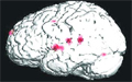
[APPLICATION OF FUNCTIONAL MR-IMAGES ACQUIRED AT LOW FIELD IN PLANNING OF NEUROSURGICAL OPERATION CLOSE TO AN ELOQUENT BRAIN AREA]
[Aim of the study - Presentation of functional MRI performed at low magnetic field (1 Tesla) for planning microsurgical operation in a patient suffering from tumor close to an eloquent brain area. Methods - Microsurgical removal navigated by frameless stereotaxy of an intrinsic tumor located in eloquent area is indicated if speech function is not damaged, i.e. exact localisation and relationship of the tumor and speech area can be defined. Before operation an optimized EPI based 2D sequence was applied to yield functional MR images. At the planning of the operation the paradigm used for the localization of the sensory language cortex contained passive listening to a text. Control investigations were performed one month postoperatively. A specific psychological test, as an additional investigation to estimate the accurate level of the sensory language function, was also conducted. Results - Low resolution (matrix of 64×64) functional MR images visualized sensory speech center and auditory cortex satisfactorily. The scans showed clearly that the Wernicke's region was situated just above the tumor (WHO grade II glioma), and this finding increased the safety of intraoperative localization and reduced the risk of morbidity. Control examinations revealed minimal decrease in sensory language function, however, it was not noticeable for either the patient or her surroundings. Conclusion - Optimized functional MR imaging performed at low magnetic field can support planning of neurosurgical operations and reduce the morbidity of microsurgical interventions.]
Clinical Neuroscience
JANUARY 20, 2007
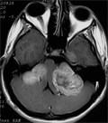
[NONSENSE MUTATION 193C>T OF NEUROFIBROMATOSIS TYPE 2 - A NEUROSURGICAL CHALLENGE]
[A 15 years old male was operated because of incidentally found intercostal schwannoma. Two years later severe cerebellar ataxy and left sided anacusis developed. MRI revealed bilateral vestibularis tumors and multiple cervical intradural extramedullar myelon compressing lesions. After partial resection of the huge left sided cerebello-pontin tumor, histologically schwannoma, and the exstirpation of the multiple cervical meningiomas the patient died three months later due to septic complications. The 24 years old mother had been operated on similar lesions 12 years earlier, after two weeks postoperative period she died. Her 14 years old twins are living, a boy also with bilateral acustic tumours and a girl who is intact. Genetic investigation revealed C>T nonsense mutation at position 193 in the exon 2 of the NF2 gene. This mutation cause premature truncation of the gene protein and is probably in connection with the clinically severe phenotype. Early diagnosis of this type of neurofibromatosis is mandatory concerning the therapy.]
Clinical Neuroscience
JANUARY 20, 2007
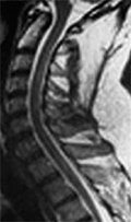
[UNCOMMON MANIFESTATION OF CENTRAL NERVOUS SYSTEM SARCOIDOSIS]
[Two cases of uncommon manifestation of central nervous system sarcoidosis are reported. A 42 year-old man had a spinal cord sarcoidosis. MRI of the spinal cord showed myelopathy in the cervico-thoracic region, and the T2-weighted image showed increasing signal intensity. Neurological symptoms did not correllate with radiological abnormalities. Neurological manifestation was paucisymptomatic. Half a year later steroid and azatioprin therapy led to almost complet radiological and clinical regression. In the second case we present a 49 year-old woman who had left side internuclear ophthalmoplegia and the brainstem lesion. The patient was proven to have sarcoidosis. In this case no abnormalities were found in brain MRI. Neurological symptoms could not be detected by MRI, probably caused by brainstem parenchymal lesions consisting of microgranulomatosis that is sarcoid "brainstem encephalitis". Neurological symptoms improved after steroid treatment in this case too. In both of the cases pulmonary lymphadenopathy helped to diagnose sarcoidosis. In our cases there were interesting correllations between neurological symptoms and MRI abnormalities. At the spinal cord sarcoidosis the radiological abnormalities were more striking than the clinical manifestation. In the other case we found distinct brainstem symptoms but could not detect MRI abnormalities.]
Clinical Neuroscience
JANUARY 20, 2007
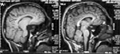
[A CASE OF SCHIZENCEPHALY WITH POLYMICROGYRIA]
[A case of extensive bilateral frontotemporal schizencephaly is alleged - more extensively in the left hemisphere - which associated with polymicrogyria. The cortical anomaly was discovered only incidentally by MR examination in a 22 year-old man who suffered from headache due to a mild head trauma. Neurological examination proved to be negative. He had no complaints or symptoms a few weeks later. The developmental anomalies in corticalisation are shortly overviewed in this group together with the possible causing factors. It has been emphasized the importance of the precise intrauterine and/or postpartum differential diagnosis between schizencephaly, porencephaly and other failure in corticalisation.]
Clinical Neuroscience
JANUARY 20, 2007

[UPDATES IN PRACTICAL NEUROLOGY - I. THE PRINCIPLES OF MODERN LEVODOPA THERAPY IN PARKINSON’S DISEASE]
[Despite the levodopa is used for the treatment of Parkinson’s disease for a long time, recently many questions raised about its clinical use. New issues emerged based on the clinical trials, on latest neuroimaging data and on better understanding the pathomechanism of motor complications. These observations have changed the routine clinical use of levodopa. In this review we summarize the evidences and practical implications of levodopa therapy.]
Clinical Neuroscience
JANUARY 20, 2007

Clinical Neuroscience
JANUARY 20, 2007

[Celebrating speech on the unveiling of the statue of academic dr. Sántha Kálmán on the 50th anniversary of his death]
[Celebrating speech on the unveiling of the statue of academic dr. Sántha Kálmán on the 50th anniversary of his death 2007;60(01-02)]
Clinical Neuroscience
JANUARY 20, 2007

[31th Congress of the Hungarian Pediatric Neurological, Neurosurgical, Pediatric and Adolescent Psychiatric Society]
[31th Congress of the Hungarian Pediatric Neurological, Neurosurgical, Pediatric and Adolescent Psychiatric Society 2007;60(01-02)]
Clinical Neuroscience
JANUARY 20, 2007

1.
Clinical Neuroscience
[Headache registry in Szeged: Experiences regarding to migraine patients]
21. MAY
2.
Clinical Neuroscience
[The new target population of stroke awareness campaign: Kindergarten students ]
21. MAY
3.
Clinical Neuroscience
Is there any difference in mortality rates of atrial fibrillation detected before or after ischemic stroke?
27. NOV
4.
Clinical Neuroscience
Factors influencing the level of stigma in Parkinson’s disease in western Turkey
27. SEP
5.
Clinical Neuroscience
[The effects of demographic and clinical factors on the severity of poststroke aphasia]
18. JUL
1.
2.
Clinical Oncology
[Pancreatic cancer: ESMO Clinical Practice Guideline for diagnosis, treatment and follow-up]
29. AUG
3.
Clinical Oncology
[Pharmacovigilance landscape – Lessons from the past and opportunities for future]
29. AUG
4.
5.