The eLitMed.hu medical portal uses computer cookies for convenient operation. Detailed information can be found in the Cookie-policy.
Clinical Neuroscience - 2006;59(05-06)
Content
Clinical Neuroscience
MAY 30, 2006
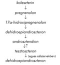
[EPILEPSY AND MALE SEXUAL DYSFUNCTION: ETIOLOGY, DIAGNOSIS AND THERAPY]
[While 10% of healthy men had sexual dysfunctions, male epilepsy patients experience sexual problems in 40-70%. The cause of sexual dysfunction in epilepsy is multifactorial, but there are three main factors: the epilepsy itself, antiepileptic treatment and psychiatrical/psychic problems. Antiepileptics with hepatic enzyme induction potential (carbamazepine, phenytoin) enhance the metabolism of sexual steroids. Valproic acid as an enzyme inhibitor and drug with high protein binding affinity elevates the free serum levels of androgenes. Certain antiepileptic drugs may have negative cognitive side effects, some of them can induce psychiatric disorders. These drugs can facilitate male sexual dysfunctions through these psychic side effects. The metabolic and endocrine alterations caused by carbamazepin may return to normal level after replacement of carbamazepin with oxcarbazepine. After an oxcarbazepin-carbamazepin replacement, carbamazepin-induced impotency can be cured. According some new data lamotrigine can also help in sexual dysfunction. The therapy of sexual dysfunction in epilepsy depends on its cause. In cases of hormonal alterations, the fist step is a change of antiepileptic regimen. Instead of enzymeinductor antiepileptics and valproate, new antiepileptic drugs should be prescribed. At present, the most investigated antiepileptic drug is the oxcarbazepine with positive effect on antiepileptic-induced male sexual dysfunction, however, lamotrigine seems to be also beneficial. If the hormonal and sexual dysfunctions cannot be eliminated by drug changes, androgenic therapy or bromocriptin may be required. Testosteron may not only be beneficial on sexual functions, but can reduce also the seizure frequency. Independent of etiology, erectile dysfunctions can be successfully treated by sildenafil.]


Clinical Neuroscience
MAY 30, 2006

[IMPORTANCE OF THE ANALYSIS OF NEUTRALIZING ANTIBODIES TO IMMUNOMODULATORY THERAPY DURING TREATMENT OF MULTIPLE SCLEROSIS]
[Interferon-α, -β, and -γ have been used for the management of several diseases with varying clinical effects. Like many other proteins, all interferon species are potentially immunogenics especially those produced by recombinant gene technologies. A reliable screening assay for anti-interferon-β antibodies is suggested for patients with multiple sclerosis receiving interferon-β therapy. Natural interferon-β is a glycosylated 166 amino acid 25 kDa protein, recombinant interferon-β is available for therapy as 1a and 1b products. Both preparations induce anti-interferon-β antibodies, detectable in the serum of interferon-β-treated patients with multiple sclerosis. The question of wich assay is optimal for testing for antiinterferon- β antibodies in interferon-β-treated patients is unsettled. Two types of antibody assays are generally used: those measuring binding antibodies and those measuring neutralizing antibodies. The findings suggest that high titers of both binding and neutralizing antibodies reduce the clinical efficacy of interferon-β in relapsing-remitting multiple sclerosis, which is important for the long-term efficacy of these drugs. Treatment with glatiramer acetat has also been shown to induce the development of “reactive antibodies” in patients with multiple sclerosis. This article briefly describes some of the findings concerning anti-interferon binding and neutralizing antibodies.]
Clinical Neuroscience
MAY 30, 2006
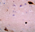
[NEUROPATHOLOGICAL EXAMINATIONS IN AUTOPSIES WITH SPECIAL FOCUS ON FINDINGS IN ALZHEIMER’S DISEASE]
[Out of an average total of 1400 autopsies per year, neuropathological examinations were performed in 477 cases between 1997 and 1999 to investigate the incidence of dementias. The majority of the studied subjects were over 50 years old. Bielschowsky's and/or Gallyas's silver methods and, in some cases, protein tau (MAP) immuncytochemistry and amyloid staining were performed beside routine examination. Pathological changes were found in 212 of the 364 cases studied by the above methods but histological changes associated with dementia were only detected in 167 cases. The various forms of Alzheimer's dementia were also classified by age. The "incipient" form of Alzheimer's disease was verified in 23 cases. Old infarcts of various extensions were found in 42 percent of Alzheimer's dementias. Very mild or age-related degenerative changes were observed in 82 cases among subjects over 50 years old. Of these, eight patients died in their 90s. In some cases (n=38) the number of neuritic plaques dominated over the number of neurofibrillary tangles but a reverse finding also occurred (n=13). Neuronal degeneration was variable and was not always proportional to the number of neurofibrillary tangles. "Simple type of senile atrophy" was defined by the presence of minor or age-related Alzheimer changes and was considered a separate entity. The "incipient" form of Alzheimer's dementia was diagnosed in relatively young individuals where mild Alzheimer changes were found at the neuropathological examination. "Preclinical" Alzheimer's dementia could only be suspected by clinical data and could very rarely be supported by the neuropathological finding of "incipient" form. The ratio of pure Alzheimer’s to vascular dementias cases proved to be 54:41 in this study. The results suggest that dementias are considerably underdiagnosed both in the clinical and pathological practice and that the recently defined "preclinical" and "incipient" forms are very hard to recognize both clinically and pathologically. The neuropathological study of the degenerative, mainly Alzheimer's type, findings in the randomly selected autopsies revealed great variations which raises many questions concerning the normal and pathologic aging of the brain as well as the "incipient" and senile forms of Alzheimer's dementia.]
Clinical Neuroscience
MAY 30, 2006

[EXPERIENCE WITH LEVETIRACETAM IN CHILDHOOD EPILEPSY]
[Objective - To evaluate the efficacy and tolerability of levetiracetam in children with drug resistant epilepsy from a retrospective study. Methods - We report the result of a study of 85 pediatric patients (mean 10.5 years, range: 1-24) with refractory generalized and focal epilepsy, who received levetiracetam as add-on treatment. The average duration of epilepsy was eight years, and the patient were treated with mean of 6.0 antiepileptic drugs befor levetiracetam was introduced. Results - Ten patients (12%) became seizuresfree, three (3%) responded with seizure reduction of more than 90%, 32 (38%) responded with seizure reduction of more than 50% following introduction of levetiracetam. No response to levetiracetam was reported in 34% (n: 29). Positive psychotopic effect was observed in 26 patient (30%). Mild to moderate side effects were reported in 11 patients (13%), consisting most frequently general behavioral changes, agression, sleep disturbances, but they ceased after decreasing the dose of levetiracetam. Mental retardation was associated with poor response and associated with more side effects. Conclusion - Levetiracetam is well tolerated new antiepileptic drug that may effectively improve seizures controll as an addon drug in resistant epilepsy in childhood with good tolerability.]
Clinical Neuroscience
MAY 30, 2006
[CHANGES IN EEG-COMPLEXITY AFTER SUBCORTICAL ISCHEMIC BRAIN DAMAGE]
[Introduction - Complexity analysis of the EEG is a relatively new field in theoretical and cinical electrophysiology. The authors present results of EEG-analysis in a patient with stroke, utilizing the sensitivity of the new procedures with respect to linear and nonlinear synchronization. Participants and methods - The EEG (19 channels) was recorded in a patient with subcortical unilateral ischaemic completed stroke involving the frontoparietal white matter while leaving the cortex intact and in 12 healthy controls in eyes open and in eyes closed conditions. Results - In the patient, increased Omega-complexity was found in slow (delta, theta) and lower alpha frequencies in the side of the stroke and in high frequencies (beta2 in eyes closed, alpha2, beta1 and beta2 in eyes open conditions) in the intact side. Synchronization likelihood was higher in the ischaemic side in the beta2 (eyes closed) and both in the beta1 and beta2 (eyes open) frequencies. Increasing Omega-complexity caused by eyes opening was markedly reduced in the patient in the beta frequencies compared to that seen in the controls. The difference was more conspicuous in the side of the infarct and involved not only the beta but also the alpha frequencies as well. Opening the eyes decreased synchronization likelihood in all frequency bands in the controls and also in the patient except the alpha2, beta1 and beta2 bands in the side of the lesion. Conclusions - The increased Omega-complexity and decreased synchronization likelihood in the slow frequencies in the infarcted side is probably the result of lesioned interneuronal connections lowering the level of cooperation of neuronal systems involved in this type of activity. The increased Omega-complexity and decreased synchronization likelihood caused by eyes opening could not be observed in the beta and alpha frequencies in the side of the lesion, possibly caused by damaged thalamocortical connections.]
Clinical Neuroscience
MAY 30, 2006
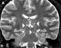
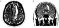
[COMPLEX GAZE DISTURBANCE CAUSED BY THALAMIC AND MESENCEPHALIC INFARCTS]
[A 36 year-old male patient developed sudden double vision and gait imbalance. Neurological examination revealed gaze paresis upward and on the left side downward (vertical “oneand- a-half”-syndrome), horizontal gaze nystagmus on the left bulbus directed to left. The MRI revealed bilateral thalamic and left midbrain ischemic lesions. The brainstem auditory and visual evoked responses were normally configured. Optokinetic nystagmus test found rightward, upward and downward hypometric saccades, convergence-retraction nystagmus - which was not visible at physical neurological examination - saccadic smooth pursuit eye movement and pseudoabducent palsy on both sides. The complex gaze disturbance was attributed to the lesions in the intralaminar nuclei of the thalamus and in the pretectal and rostromedial tegmentum of the mesencephalon. Infarcts may have been due to a variant artery: i.e. the thalamoperforant and the superior paramedian mesencephalic arteries originate with common branch from one of the communicant basilar artery. The authors discuss the mechanism of complex gaze palsy and call attention to the diagnostic value of optokinetic nystagmus examination.]
Clinical Neuroscience
MAY 30, 2006

[ABSTRACTS OF THE 8TH CONGRESS OF THE HUNGARIAN EPILEPSY LEAGUE Pécs, 25-27 May, 2006.]
[Abstracts of the 8th congress of the hungarian epilepsy league Pecs 25-27 may 2006]
Clinical Neuroscience
MAY 30, 2006

[INITIAL STAGES OF ANTHROPOLOGY IN HUNGARY]
[In the second half of the 19th century anthropological researches started everywhere in the world. Cranioscopy formed an important part of the biological anthropology. József Lenhossék (1818-1888) worked also on this subject and on the basis of one of his researches in 1875 he became the founder of the anthropology in Hungary. On 76 skulls of several collections and on 265 heads together with his coworkers he performed 50 measurements on each skulls and heads and calculated the important ratios (skull-indexes). He determined the skull-indexes of the Hungarian people. These indexes are valid also today.]
Clinical Neuroscience
MAY 30, 2006

Clinical Neuroscience
MAY 30, 2006
Clinical Neuroscience
MAY 30, 2006

Clinical Neuroscience
MAY 30, 2006

1.
Clinical Neuroscience
[Headache registry in Szeged: Experiences regarding to migraine patients]
21. MAY
2.
Clinical Neuroscience
[The new target population of stroke awareness campaign: Kindergarten students ]
21. MAY
3.
Clinical Neuroscience
Is there any difference in mortality rates of atrial fibrillation detected before or after ischemic stroke?
27. NOV
4.
Clinical Neuroscience
Factors influencing the level of stigma in Parkinson’s disease in western Turkey
27. SEP
5.
Clinical Neuroscience
[The effects of demographic and clinical factors on the severity of poststroke aphasia]
18. JUL
1.
2.
Clinical Oncology
[Pancreatic cancer: ESMO Clinical Practice Guideline for diagnosis, treatment and follow-up]
29. AUG
3.
Clinical Oncology
[Pharmacovigilance landscape – Lessons from the past and opportunities for future]
29. AUG
4.
5.