The eLitMed.hu medical portal uses computer cookies for convenient operation. Detailed information can be found in the Cookie-policy.
Clinical Neuroscience - 2003;56(07-08)
Content
Clinical Neuroscience
AUGUST 20, 2003

[Apoptosis in focal brain ischaemia]
[Ischaemic stroke is one of the major causes of death and disability in the developed world. It is caused by focal impairment of cerebral blood flow. The subsequent ischaemic cell death is predominantly necrotic in nature. However, a therapeutically important characteristic is the delayed apoptotic cell demise in the border zone of the primary lesion core. Apoptosis is one of the most intensively studied field of current medical and biological research. The better understanding of its mechanism may provide novel and more effective ways of therapy in a wide range of diseases including ischemic stroke. The salient neurological features of focal brain ischaemia and the morphological signs of apoptotic and necrotic cell death are summarized. The mechanism of apoptosis is discussed. It is divided into an early genetic phase of decisionmaking followed by a cellular execution phase. The characteristics of the early shift in the finely tuned balance of proand antiapoptotic genes and their protein products, which is preceded by an inbalance in intracellular ionized calcium homeostasis, energy depletion and mitochondrial dysfunction is discussed. The crucial role of caspases in apoptosis is emphasized. The three possible pathways during the execution phase is described: the intrinsic- and extrinsic caspase activation cascade and the caspase-independent intracellular signal transduction route. The molecular mechanism of neural cell membrane damage in the execution phase is discussed and some examples of altered protein synthesis also known as message-selection are given. The important role of late reperfusion in the execution phase is emphasized. The possible targets of antiapoptotic therapeutic approaches and the results of experimental studies are presented as well as the perspectives of their use in human clinical care.]


Clinical Neuroscience
AUGUST 20, 2003
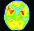
[Imaging of dopamine transporter with 99mTc-TRODAT-SPECT in movement disorders]
[99mTc-TRODAT-1 is a new, technetium based radiopharmaceutical that selectively binds to the dopamine transporters. The aim of the study was to evaluate the dopamine transporter status in movement disorders. Methods - In eight healthy volunteers (age range 22-58 years), 28 patients with Parkinson’s disease (age range 42-80 years), 10 patients with Parkinsonian syndrome (age range 51-79 years) and 13 patients with essential tremor (age range 43-71 years) were 99mTc-TRODAT-SPECT tests performed. The results were evaluated visually and semiquantitatively. Results - The visual assessments were concordant with those of the semiquantitative in each case. The 99mTc- TRODAT uptake of the striatum was referenced to the cerebellum, the frontal and occipital cortex. The best deviation was found in aspect of the occipital cortex. The striatum/occipital ratio was the following: healthy volunteers: 2.12±0.27; Parkinson’s disease: 1.52±0.27; Parkinsonian syndrome: 1.57±0.26; essential tremor: 2.06±0.69. The striatal dopamine transporter availability was significantly lower in subjects with Parkinson's disease or Parkinsonian syndrome compared to the control subjects. There was no difference between healthy volunteers and patients with essential tremor. Using discriminant analysis, the discriminant function had significantly different values in the group of Parkinson’s disease than in Parkinsonian syndrome: f= -3.675×caud/occipit+6.293×put/occipit -2.548. Conclusion - 99mTc-TRODAT-SPECT is able to visualise the presynaptic dopaminergic degeneration. This method itself can be useful in differential diagnosis in some type of movement disorders.]
Clinical Neuroscience
AUGUST 20, 2003
[Two cases of frontotemporal dementia]
[Frontotemporal dementias represent the third most common cause of primer degenerative dementias next to Alzheimer’s disease and Lewy body disease. Frontotemporal dementia constitutes 10-20% of all praesenilis dementias. The authors present the results of the 10 years' clinical, neuropsychological, neuropathological examinations and brain imaging with the examples of two cases. At the early stage of frontotemporal dementia changes of personality and social conduct are prominent, whereas cognitive functions are relativelly well preserved. The usual dementia tests are not sufficiently sensitive to disclose noncognitive symptoms. Clinical diagnosis as well as differentiation from functional psychiatric disorders can be difficult. Brain imaging present the frontal and the anterior temporal lobe atrophy and selective hypometabolism in these areas. The typical onset is between at the age 50 and 65 years. It is very rare under the age of 30. The symptoms of two patients started at the age of 42-44. The first diagnosis was post traumatic stress disorder. Later stereotyped behaviour, mental rigidity, hyperorality, irritability, progressive reduction of speech and vegetative dysfunctions appeared. Besides the affecting of the irresistibly worsening symptoms and the medical care requiring strength and inventiveness, the authentic informing of the relatives is also a challenge. The caregivers have special relationship with the patients and their relatives.]
Clinical Neuroscience
AUGUST 20, 2003

[Positron emission tomography in presurgical localization of epileptic foci]
[The success of cortical resection for intractable epilepsy of neocortical origin is highly dependent on the accurate presurgical delineation of the regions responsible for generating seizures. In addition to EEG and structural imaging studies, functional neuroimaging such as positron emission tomography (PET) can assist lateralization and localization of epileptogenic cortical areas. In the presented studies, objectively delineated focal PET abnormalities have been analyzed in patients (mostly children) with intractable epilepsy, using two different tracers: 2-deoxy-2-[18F]fluoro-D-glucose (FDG), that measures regional brain glucose metabolism, and [11C]flumazenil (FMZ), that binds to GABAA receptors. The PET abnormalities were correlated with scalp and intracranial EEG findings, structural brain abnormalities, as well as surgical outcome data. In patients with extratemporal foci and no lesion on MRI, FMZ PET was more sensitive than FDG PET for identification of the seizure onset zone defined by intracranial EEG monitoring. In contrast, seizures commonly originated from the border of hypometabolic cortex detected by FDG PET suggesting that such areas are most likely epileptogenic, and should be addressed if subdural EEG is applied to delineate epileptic cortex. In patients with cortical lesions, perilesional cortex with decreased FMZ binding was significantly smaller than corresponding areas of glucose hypometabolism, and correlated well with spiking cortex. Extent of perilesional hypometabolism, on the other hand, showed a correlation with the life-time number of seizures suggesting a seizurerelated progression of brain dysfunction. FMZ PET proved to be also very sensitive for detection of dual pathology (coexistence of an epileptogenic cortical lesion and hippocampal sclerosis). This has a major clinical importance since resection of both the cortical lesion and the atrophic hippocampus is required to achieve optimal surgical results. Finally, the author demonstrated that in patients with neocortical epilepsy, FDG PET abnormalities correctly regionalize the epileptogenic area, but their size is not related to the extent of epileptogenic tissue to be removed. In contrast, complete resection of cortex with decreased FMZ binding predicts good surgical outcome suggesting that application of FMZ PET can improve surgical results in selected patients with intractable epilepsy of neocortical origin.]
Clinical Neuroscience
AUGUST 20, 2003

Clinical Neuroscience
AUGUST 20, 2003

Clinical Neuroscience
AUGUST 20, 2003

[6th National Congress of the Hungarian Stroke Society]
[6th National Congress of the Hungarian Stroke Society 2003;56(07-08)]
Clinical Neuroscience
AUGUST 20, 2003

1.
Clinical Neuroscience
[Headache registry in Szeged: Experiences regarding to migraine patients]
21. MAY
2.
Clinical Neuroscience
[The new target population of stroke awareness campaign: Kindergarten students ]
21. MAY
3.
Clinical Neuroscience
Is there any difference in mortality rates of atrial fibrillation detected before or after ischemic stroke?
27. NOV
4.
Clinical Neuroscience
Factors influencing the level of stigma in Parkinson’s disease in western Turkey
27. SEP
5.
Clinical Neuroscience
[The effects of demographic and clinical factors on the severity of poststroke aphasia]
18. JUL
1.
2.
Clinical Oncology
[Pancreatic cancer: ESMO Clinical Practice Guideline for diagnosis, treatment and follow-up]
29. AUG
3.
Clinical Oncology
[Pharmacovigilance landscape – Lessons from the past and opportunities for future]
29. AUG
4.
5.