The eLitMed.hu medical portal uses computer cookies for convenient operation. Detailed information can be found in the Cookie-policy.
Clinical Neuroscience - 2003;56(03-04)
Content
Clinical Neuroscience
APRIL 20, 2003
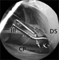
[Biportal neuroendoscopy of the prepontine cisterns]
[Introduction - While bi- or multiportal approaches have been adopted in different fields of surgery including abdominal and spine surgery, the uniportal access into the skull is a traditional principle in neurosurgery. In this preclinical cadaver study the authors developed combinations of biportal endoneurosurgical dissections in the prepontine subarachnoid space to test the safety of this technique. Methods - In 34 fresh post-mortem adult human cadavers and 14 formaldehyde-fixed adult human head specimen a total of 48 biportal endoscopical dissections were carried out. 0°, 30°, and 70° lens scopes with a diameter of 1.7 and 4.2 mm and trochars with a diameter of 5.0 to 6.5 mm were used. Results - Six different endoscopic routes to the prepontine region and a total of 10 different combinations of this approaches could be described. Useful and safe biportal combinations were: 1. supraorbital on both sides, 2. supraorbital combined with ipsilateral anterior subtemporal, 3. supraorbital combined with contralateral anterior subtemporal, 4. supraorbital combined with ipsilateral posterior subtemporal, 5. supraorbital combined with ipsilateral frontal interhemispheric, 6. supraorbital combined with contralateral frontal interhemispheric, 7. anterior subtemporal combined with ipsilateral frontal interhemispheric, 8. anterior subtemporal combined with contralateral frontal interhemispheric. Conclusion - The biportal endomicrosurgical strategy offered effective and safe dissections within the prepontine subarachnoid space.]


Clinical Neuroscience
APRIL 20, 2003

[Neuropsychological applications of the Rey Complex Figure and Recognition Test]
[The authors describe the clinical applications of the Rey Complex Figure and Recognition Test and explain how the individual abilities and information-processing characteristics are reflected. Sequential and quantitative evaluation methods are presented. Summarizing the relation between IQ and RCFT they evaluate the differencies in task completion strategies of healthy subjects and subjects with brain damage, suggesting that these differences originate in changes of the information-processing capacity. Application of the RCFT in clinical child psychology is also suggested, primarily in differential diagnostics of children with attention disorder and disruptive disorder. The authors illustrate the protocols of the neuropsychological examination and the role of the RCFT with a case study.]
Clinical Neuroscience
APRIL 20, 2003

[Occipital spectral EEG-parameters in newly diagnosed, untreated epilepsy patients]
[Introduction - Minor spectral EEG alterations hidden to the naked eye may be of interest in the field of epileptology, cognitive performances, and drug effects. In order to introduce new scientific results of brain wave research into the clinical field of epilepsy- and drug-related cognitive problems, a normative quantitative EEG database for epilepsy was constructed. Patients and methods - 171 newly referred, five to 50 years old patients with untreated ”new” epilepsy (that is, clinical, EEG, MRI investigations had been done in 24 months after the first unprovoked seizure) were collected. EEG was recorded with closed eyes, in the waking-relaxed state. Effects that are known to influence EEG spectra (nearby seizures, drugs, etc.) were excluded as far as possible. A total of two minutes of waking-resting EEG activity was chosen for spectral analysis. Fast Fourier transformation of the selected samples were calculated resulting in absolute power, percent power and mean alpha frequency (AA, RA, and AMF respectively) for the right and left occipital derivations. For each patient (and also for 37 healthy controls), the deviation of the individual values from the age-adjusted normative mean was expressed in Z-score. Main diagnostic epilepsy categories were compared to the control group as well as to each other. In addition, effects of MRI-defined cerebral lesions and interictal spiking on spectral EEG parameters were investigated. Results - All group averages were within the 95 per cent confidence interval. Overwhelming majority of the individual data fell within a 3Z range. Statistically significant differences were found for AA and RA, but seldom for AMF. Right and left alpha-parameters were surprisingly symmetrical in all groups. The main difference between epilepsy groups and controls was less AA and RA power in the epilepsy groups. MRI-defined lesions and interictal epileptiform activity did not significantly influence EEG spectral variables. Conclusion - These results might serve as reference data and might help planning of further quantitative EEG studies in the triangle of epilepsy, cognitive problems, and drug effects.]
Clinical Neuroscience
APRIL 20, 2003
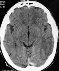
[Cerebral sinusthrombosis and ulcerative colitis - two cases]
[Inflammatory bowel diseases (IBD) - ulcerative colitis and Crohn’s disease - are associated with increased risk for thrombotic complications both in the arterial and venous system. Cerebral sinus thrombosis is a rare but potentially fatal consequence of these diseases. Modern imaging methods made this uncommon complication of IBD more frequently recognized. The link between IBDs and thrombosis has been extensively studied. Inherited coagulation disorders (APC resistance, antithrombin III and protein-S deficiency), acquired diseases (antiphospholipid syndrome), and the frequent use of corticosteroids were suspected. Two cases of ulcerative colitis associated with cerebral sinusthrombosis successfully treated are reported. The connection between IBD and thrombotic complications and the therapeutic risks are discussed as well.]
Clinical Neuroscience
APRIL 20, 2003
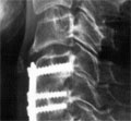
[Surgery of ventral intradural midline cervical spinal pathologies via anterior cervical approach: our experience]
[Introduction - The surgical removal of the cervical intradural pathologies located ventrally carries a high risk. According to the anatomical situation and the increasing experience with anterior cervical approach and corpectomy revealed the reality to remove the ventral midline pathologies this way. The anterior approach which require corpectomy preferable to cervical intradural lesions located ventrally at the midline. In the literature have described anterior approach for intradural cervical lesions in very limited cases. Case - The authors present five cases of intradural ventral cervical spinal pathologies, where removal was done via anterior cervical approach with corpectomy. Two of the cases were intradural meningeomas, one intramedullary cavernoma, one ventral arachnoid cyst and one malignant neurogenic tumour. The approach was described elsewhere. The corpectomy gave a relatively wide window to explore the pathologies and under operative microscope the local control of removal was fairly well. After the total removal of tumours and cavernoma, and fenestration of arachnoid cyst to the subarachnoid space watertight dural closure was made and the cervical spine was stabilized with autolog iliac bone graft, plate and screws. The recovery of the patients was well and there were no postoperative complications. Conclusions - The anterior cervical approach with corpectomy seems to be a real and safe way to explore and remove the cervical ventral midline pathologies. Postoperative MRI has a great value in early control after the surgery and for follow up the patients.]
Clinical Neuroscience
APRIL 20, 2003

Clinical Neuroscience
APRIL 20, 2003

Clinical Neuroscience
APRIL 20, 2003

Clinical Neuroscience
APRIL 20, 2003

Clinical Neuroscience
APRIL 20, 2003

Clinical Neuroscience
APRIL 20, 2003

1.
Clinical Neuroscience
[Headache registry in Szeged: Experiences regarding to migraine patients]
21. MAY
2.
Clinical Neuroscience
[The new target population of stroke awareness campaign: Kindergarten students ]
21. MAY
3.
Clinical Neuroscience
Is there any difference in mortality rates of atrial fibrillation detected before or after ischemic stroke?
27. NOV
4.
Clinical Neuroscience
Factors influencing the level of stigma in Parkinson’s disease in western Turkey
27. SEP
5.
Clinical Neuroscience
[The effects of demographic and clinical factors on the severity of poststroke aphasia]
18. JUL
1.
2.
Clinical Oncology
[Pancreatic cancer: ESMO Clinical Practice Guideline for diagnosis, treatment and follow-up]
29. AUG
3.
Clinical Oncology
[Pharmacovigilance landscape – Lessons from the past and opportunities for future]
29. AUG
4.
5.